Category: Diagnostic Physics

30 Jun 2023
Diagnostic and Radiation Therapy Examples

Other Forms of Radiation Therapy and Diagnostic Machines

In our last blog post, we explored the timeline of diagnostic imaging and radiotherapy developed using x-rays. It was the original discovery of x-rays by Röntgen that led to the creation of these medical practices. However, other researchers of the 20th century also invented diagnostic tools and radiation therapy without x-ray involvement at all. We will explore a few of these imaging and radiotherapy machines or procedures in this blog.

Diagnostic Imaging

MRI

One of the most well-known imaging procedures is MRI, which stands for Magnetic Resonance Imaging. Research for MRI began early in the 1970s, but investigation of the magnetic resonance principles started as early as 1945. By happenstance, Felix Bloch, a Swiss physicist working at Stanford, and American physicist, Edward Mills Purcell at Harvard, conducted—nearly simultaneously—an experiment for a new mode of nuclear induction. This led to published papers from both physicists on nuclear magnetic resonance. After publication, both Purcell and Bloch won the 1952 Nobel Prize for this research.1

The process to produce 2D images in an MRI using nuclear magnetic resonance (NMR) was discovered by Paul Lauterbur and Peter Mansfield. Lauterbur published the first nuclear magnetic image in 1973, eventually producing 3D images as well. However, he still had long to go before these images would be practical for application with actual patients. Mansfield continued to expand on Lauterbur’s work, developing the echo-planar imaging technique to improve image quality and scan time in the late 1970s.  By 1977, physician Raymond Damadian created the first full body MRI machine. Through continuous improvements and further research, today’s MRI provides a safe, effective way of capturing images in the brain, portions of the body, the cardiovascular system, and central nervous system. MRIs provide information on size, location, classification, and grade of lesions so as to aid diagnosing stroke, MS, blood supply issues, and any brain damage.2

Ultrasound

Today, ultrasound is most widely known in the OB/GYN field of medicine. Through its invention, however, this diagnostic tool proved its range of utility before becoming a staple in wellness checks during pregnancies. The origin of ultrasonography is often credited to Lazzaro Spallananzi, a physiologist who discovered echolocation throughout 1794 from various experiments involving bats. The principles that serve as a basis for echolocation are also those functioning for medical ultrasound technology today. In 1942, neurologist Karl Dussik first used ultrasonic waves in a diagnostic procedure while attempting to find brain tumors. He later published a report of musculoskeletal ultrasonography in 1958, laying the groundwork for diagnostic musculoskeletal ultrasound.3

Professor Ian Donald from the University of Glasgow was responsible for the development of medical ultrasound in clinical practice. During his research from the late 1950s into the 1960s, there was clinical skepticism about the use of ultrasound. Many doctors felt that manual abdominal and pelvic examinations had proven adequate for medical diagnoses. With his co-workers, Donald performed several studies to show the likelihood of misdiagnoses of a cyst versus a malignant mass. By publishing the findings in 1958, this became a critical point in time for encouraging use of the medical ultrasound. Donald and his colleagues eventually developed an automatic scanner that utilized a full bladder for early pregnancy detection in 1963. This set the path to the now common practice of ultrasounds in the OB/GYN field today.4

PET Scans

Experimental physicist, C.D. Anderson, observed particles from cosmic rays in 1932 that were the same mass as an electron, but which moved in a strong magnetic field along a path opposite to that of an electron indicating a positive charge. He named these particles as “positrons” or positive electrons.

Emission of these positrons was the proof that Irène Joliot-Curie and Frédéric Joliot-Curie used to prove the artificial creation of radioactive elements to win the 1935 Nobel Prize in Chemistry. The creation of these artificial elements was the main focus over the following years due to their significance to military research from the late 1930s until the 1950s. Imaging studies began taking strides as a result of the desire by Louis Sokoloff, an American neuroscientist who hoped to relate mental function to brain function. This encouraged research through the 1950s until the first tomography unit attempt in 1968. This was the turning point in which PET scan development accelerated into the machines we are more familiar with today.5

PET scans can provide imaging through the detection of the gamma ray pairs that are created when a positron annihilates with an electron inside the tissue where the positron emitting isotope has collected. Whereas other diagnostic scans capture the form of an object, PET scans correlate with the metabolic functions of the tissues that uptake the radioactive compound. The compound with the radioactive tracer is typically a form of glucose called FDG (fluorodeoxyglucose), which is used as an energy source by cells and is given to the patient by means of an IV injection. The more rapid the metabolism of the cells in a tissue, as you have with cancer cells, the more uptake of the FDG. This differential uptake is then seen as an enhanced region on a PET scan. 5

Radiation Therapy

Gamma Rays – Gamma Knife (and Cobalt Therapy)

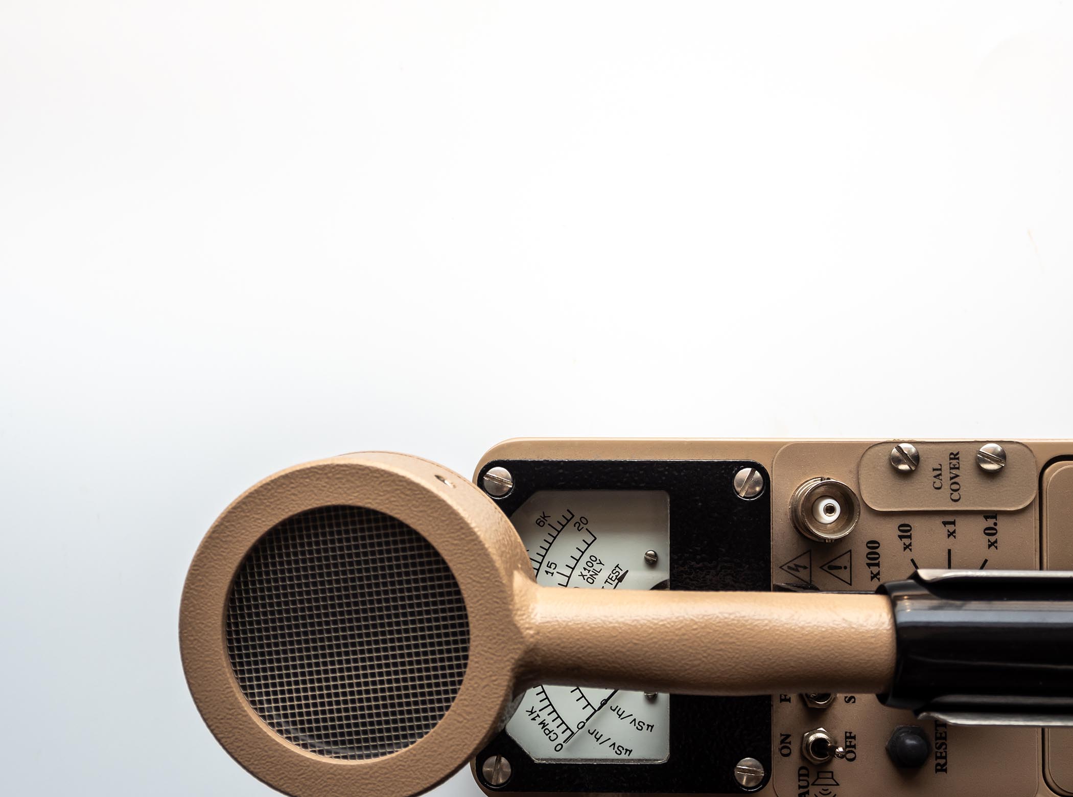
Gamma radiation from radioactive cobalt sources has been harnessed to create an alternative method for producing therapeutic radiation beams. Similar in design to the previously discussed LINAC machines, a Cobalt unit uses a single Co-60 source to produce the radiation beam for treatment. A machine of a fundamentally different design, the Gamma Knife was developed for the treatment of brain tumors. The Gamma Knife is a stereotactic radiosurgery technique that allows for functional neurological surgery. This machine uses anywhere from 192 to 201 C0-60 arrayed in a semi-spherical shield with each source collimated to point their radiation to the same point in space. This type of precision allows for the potential to treat brain tumors and, in turn, pain, movement disorders, and even some behavioral disorders for patients who had not been responsive to other treatment.6

The Gamma Knife was created through the inspirations of Swedish professors Borje Larsson and Larks Leksell in the 1950s. After an investigation using proton beams with stereotactic devices capable of pinpointing targets in the brain, the researchers eventually gave up on that approach due to cost and complexity. Motivated to find an alternative, Larsson and Leksell went on to create a prototype Gamma Knife system in 1967. The prototype unit was a success, used in Sweden for twelve years  and led to a second Gamma Knife in 1975, before other units began appearing globally in the 1980s.6

Proton Therapy

Protons were discovered in the early 1900s. New Zealand physicist Ernest Rutherford’s research during this time led to his discovery of a nuclear reaction that led to the “splitting” of an atom, where he found protons. As a charged particle, the protons have a finite range in matter. The proton’s interaction with matter produces ionization as the proton slows down along its path, losing energy. A peak of deposited dose then occurs at a depth that is proportional to the proton’s original energy.7

The cyclotron, invented by the American physicist Ernest O. Lawrence along with his associates in 1929, proved an efficient way to produce beams of protons. The cyclotron was capable of accelerating protons to high enough energy that, towards the middle of the 20th century, it was suitable for application in cancer treatments.

Dr. Robert Wilson, another American physicist, was the one to spark the idea of proton therapy for battling cancer. He wrote a seminal paper on the functionality of particle beams (comprised of either protons or other heavy-charged particles) and their ability to disperse their energy in the body—the initial release would be small, then exponentially grow when the beam reaches the end of its path for maximum effect against the tumor. Dr. Wilson published the paper in 1946 and received credit to being the inspiration of further research on proton therapy and the continued development of cyclotrons.7

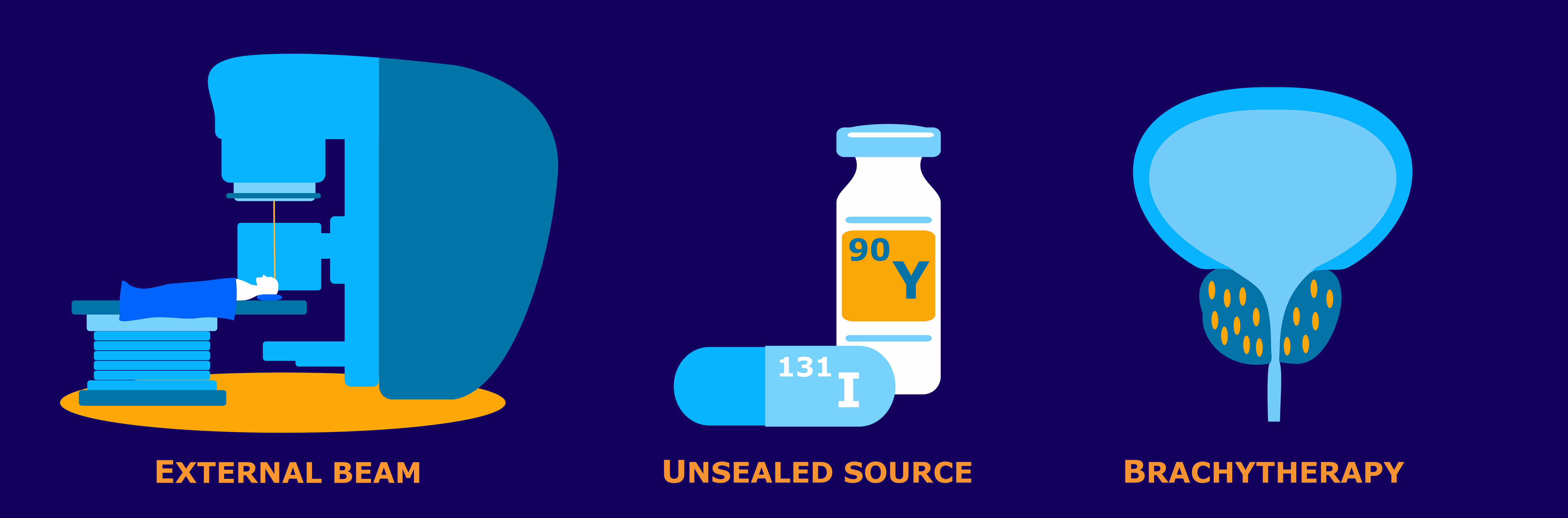
Brachytherapy and Interstitial Therapy

Brachytherapy is an internal radiation therapy technique involving direct placement of radiation inside a patient’s body cavity. This procedure involves small, radioactive source implants that are generally positioned in a body cavity to be near a cancerous tumor. The radioactive material, typically encapsulated within suitable housing material such as titanium, constantly exposes the tumor to a stream of radiation until removal of the radioactivity. These implants will be temporary, and the patient is either hospitalized or kept in a special suite for the duration of the treatment.

Another form of therapy, interstitial therapy (or interstitial brachytherapy), uses sources that are placed directly into the tissue. These may either be temporary or permanent implants of small, encapsulated sources on the order of the size of a rice grain. For treatments with permanent implants, it is safe enough for the patient to go home with a few simple safety guidelines to follow.8

Brachytherapy was first used in 1901 during an attempt to treat lupus. Alexandre Danlos and Paul Bloch completed this treatment with a radioactive sample from Marie Curie. Shortly after, in 1903, Margareth Cleaves used brachytherapy to treat cervical cancer. It became a popular radiotherapy technique to treat breast, cervical, and prostate cancer. As technology advanced along with the use of brachytherapy through the 1900s, imaging-guidance became a valuable asset. More precise dosimetry became possible and allowed for better brachytherapy planning when doctors could use diagnostic modalities such as CTs, MRIs, or ultrasounds. Today, brachytherapy is a more popular treatment for conditions ranging through gynecological, genitourinary, ocular, and head & neck cancers.9

Summary and Conclusion

The 20th century saw tremendous growth and development in diagnostic machinery and radiotherapy. From soundwave technology that gave way to ultrasound, magnetic resonance inspiring MRIs, and the discovery of positrons, medical and non-medical fields can run diagnostic imaging that suits their needs exactly for the most effective information. Radiation therapy research continuously improves the technology and practices used for the wellbeing of patients. Precision instruments such as the Gamma Knife, Cyberknife, and modern LINACs permit for types of treatments that would otherwise be impossible whilst at the same time reducing the side effects associated with external beam therapy. Proton therapy calculations allow for precise travel through the body for maximum effect against cancerous tumors. Brachytherapy provides a treatment option that directly targets tumors without having radiation travel through the body to the target tissue. Modern image guidance ensures source placement exactly where treatment is necessary and that externally directed therapy beams hit their target. As these modalities evolve, we witness the growing reality of safer, more effective diagnoses and cancer treatments within our lifetimes.

The Versant Physics team has experience that covers a range of equipment. This includes dental units, mobile c-arms and Cone-beam CTs, as well as high energy LINACs and even Proton Therapy units and Cyclotrons. To learn more about how our services can help you, contact us to set up a meeting.

Sources

1. NMR Basics | Nuclear Magnetic Resonance Spectroscopy Facility | University of Colorado Boulder. www.colorado.edu. https://www.colorado.edu/lab/nmr/nmr-basics

2. The History of the MRI – DirectMed Parts & Service. https://directmedparts.com/history-of-the-mri/

3. D. Kane and others, A brief history of musculoskeletal ultrasound: ‘From bats and ships to babies and hips’, Rheumatology, Volume 43, Issue 7, July 2004, Pages 931–933, https://doi.org/10.1093/rheumatology/keh004

4. History of Ultrasound – Overview of Sonography History and Discovery. Ultrasoundschoolsinfo.com. Published December 27, 2021. https://www.ultrasoundschoolsinfo.com/history/

5. Henry N. Wagner, A brief history of positron emission tomography (PET), Seminars in Nuclear Medicine, Volume 28, Issue 3, 1998, Pages 213-220, ISSN 0001-2998, https://doi.org/10.1016/S0001-2998(98)80027-

6. History and Technical Overview | Neurosurgery. Neurosurgery. Published 2018. https://med.virginia.edu/neurosurgery/services/gamma-knife/for-physicians/history-and-technical-overview/

7. History of Proton Therapy – NAPT. NAPT. Published 2018. https://www.proton-therapy.org/about/history-of-proton-therapy/

8. Radiation Oncology. Radiation Answers. Accessed May 31, 2023. https://www.radiationanswers.org/radiation-sources-uses/medical-uses/radiation-oncology.html

9. Mayer C, Kumar A. Brachytherapy. PubMed. Published 2021. https://www.ncbi.nlm.nih.gov/books/NBK562190/

01 Jun 2022
Patient Wearing Head Coil During MRI Scan In Hospital

Diagnostic Medical Physics in Medicine: Why It’s Important

Many are unfamiliar with the important role that diagnostic medical physics plays in medicine, particularly in the diagnosis and treatment of diseases like cancer.

At Versant Physics, we provide a wide range of diagnostic medical physics services that help healthcare facilities safely and effectively execute procedures for the health and well-being of their patients. Our goal is to help facilities ensure their patients are protected from excessive levels of radiation and that diagnostic equipment is working appropriately, all while maintaining compliance with state and federal regulations.

In this blog post, we’ll break down what diagnostic imaging is, how and why physics principles are applied to diagnostic medicine, and the various roles of a diagnostic medical physicist to help clarify the importance of this profession.

What is Diagnostic Imaging?

Diagnostic Imaging is a range of techniques and equipment used to look inside the body. The purpose of this is to help physicians identify injuries and illnesses, and to help make an accurate diagnosis and treatment plan. This can include a variety of procedures, from simple X-rays for broken bones to more complex procedures involving the brain, heart, or lungs.

Diagnostic imaging procedures are usually painless and noninvasive. However, depending on the test being performed, some patients may be exposed to small amounts of radiation.

diagnostic medical physics

CT scans are a common example of a diagnostic imaging test that emits radiation. In a CT scan, the patient is exposed to a series of X-rays from a variety of angles which are then processed via a computer. The computer creates cross-sectional images of the inside of the body. CT scans are higher-quality images than a normal X-ray and allow physicians to view both hard and soft tissues in the body. They can check for stroke, internal bleeding, chest abnormalities, enlarged lymph nodes, abdominal or pelvic pain, tumors, and more. It is also used to monitor existing diseases such as heart disease and cancer. 

Other common diagnostic imaging procedures include mammography, which helps detect and diagnose breast cancer, fluoroscopy, magnetic resonance imaging (MRI), and ultrasounds.  

Diagnostic Physics and Medicine

Medical physics as a field is divided into five categories, including:

  • nuclear medicine
  • therapeutic medical physics,
  • medical health physics,
  • magnetic resonance imaging physics, and
  • diagnostic imaging.

Diagnostic medical physicists are responsible for ensuring the safe and effective application of radiation used in medical treatments. Specifically, radiology procedures. They work as a member of a patient’s care team, which typically includes physicians, dosimetrists, and radiologic technologists among others.

Equipment Evaluation and Compliance

One of the main roles of a diagnostic medical physicist is to ensure the safe operation of radiation-producing machines and diagnostic radiation detectors. This can include developing imaging equipment specifications, measuring the radiation produced by a piece of equipment prior to clinical use, and proving that the equipment is compliant with regulatory and accreditation requirements.

This also includes assessing all the software, algorithms, data, and computer systems associated with the radiation-producing equipment for accuracy and performance.

Acceptance Testing

Any unit that is used in a diagnostic setting must be periodically reviewed to ensure not only that the image quality is maintained, but that the unit is operating in compliance with the manufacturer’s specifications.

Most states require that a newly installed piece of diagnostic imaging equipment, whether it is brand new or used, be tested by a qualified medical physicist prior to first clinical use. This extremely thorough survey confirms that the unit was installed and set up correctly and ensures that it meets vendor and industry performance standards. It is also an opportunity to identify any potential issues with the unit before it is used on patients.

mammography unit

Typical units that require acceptance testing include fluoroscopic x-rays, radiographic x-rays, PET and PET/CT units, mammography equipment, C-arms, CTs, SPECT cameras, and PACS workstations.

Commissioning

The commissioning process for diagnostic radiation therapy machines such as Linear Accelerators involves testing the unit’s functionality and verifying that dose calculation algorithms work appropriately to produce measured dose calculations.  

Radiation-producing equipment like a LINAC is highly technical and specific. There are many requirements and protocols that detail how this unit should work, from how much energy it produces to the shape and direction of the beam. Diagnostic medical physicists are trained to measure, assess, and implement the optimal baseline values for a unit during the commissioning process.

Patient safety is the end goal of all diagnostic physics commissioning work.

Shielding

Another important aspect of diagnostic physics includes the planning and placement of shielding in areas that use radiation. In the United States, 35+ states require specific shielding designs in any room that houses radiation-producing equipment.

A diagnostic medical physicist can evaluate any shielding that is installed to determine if it will adequately protect workers, patients, and the public from the radiation outside of the scope of a specific treatment. This includes planning for material thickness as well as appropriate placement.

Versant Physics physicists are experienced with a range of equipment shielding requirements, including dental units, Cone-beam CTs, mobile c-arms, high-energy LINACS, Proton Therapy units, and Cyclotrons.

Our team is also experienced with different types of shielding materials, including non-lead materials, which are guaranteed to meet regulatory guidelines and ALARA principles.

Patient Dose & Treatment

Part of a diagnostic physicist’s job is also to ensure the safety of medical imaging modalities being applied in the treatment of individual patients.

They are responsible for determining the exact radiation dose a patient will receive in accordance with the radiation oncologist’s prescription before the patient begins treatment. Creating this therapy plan can take a few hours or multiple days, depending on the complexity of the illness. They also ensure radiation protection guidelines are in place, develop QA tools that ensure optimal image quality, and make sure that all operators are trained in the use of the best imaging techniques.

A diagnostic medical physicist may also monitor the dose of the patient throughout the course of their treatment.

Patients rarely interact directly with the medical physicist on their care team; however, they are a vital part of a safe and effective treatment process.

Versant Physics Diagnostic Support

Our board-certified physicists are able to handle diagnostic physics support for a variety of facilities, including hospitals, clinics, dental offices, and university health systems. With decades of experience, top-of-the-line equipment, and a passion for patient safety, our team is the best choice to assist with your diagnostic medical physics needs.

Contact us for a quote or to learn more about our medical physics support services.

28 Apr 2022
Radiation Protection Survey of Package with Pancake Probe

A Beginner’s Guide to Radiation Protection Surveys

The purpose of a radiation protection survey is to identify higher-than-normal doses of radiation in medical environments, labs, and anywhere radiation-emitting machines or radioactive materials (RAM) are used. They are required by state and federal regulations to be performed regularly to ensure the safety of technicians, technologists, nurses, doctors, researchers, and patients.

In this brief guide we’ll talk about what a radiation protection survey is, why it is important, and the type of equipment required to perform a radiation protection survey.

What is a radiation protection survey?

Radiation protection surveys are a way to directly measure radiation levels and identify potential leakage through breaks or voids in shielding.

Surveys are performed on:

  • Diagnostic fluoroscopic and radiographic equipment
  • Non-medical industrial equipment such as those found in veterinary offices
  • CT and CBCT machines
  • Particle accelerators
  • Irradiators
  • Bone mineral densitometers
  • Cabinet x-ray machines
  • Areas that use sealed sources of RAM
  • Packages containing RAM

The Different Types of Radiation Surveys

Not all radiation surveys are created equal. Let’s talk about some of the different surveys you may encounter a need for in your radiation safety program.

Radiation Emitting Device Survey

X-ray machines and other radiation-emitting devices require regular surveys to be performed to confirm that the machine is operating as expected. Radiation producing machines are surveyed for:

  • Timer accuracy
  • Radiation output
  • Focal spot size
  • kVp and mA
  • Beam limitation accuracy
  • Filtration
  • Skin entrance exposure / rate of exposure
  • Scatter radiation measurements
  • Photo-timer operation
  • Proper signage, labels, and postings

If high or unexpected dose rates are measured during a survey, the machine should be turned off and undergo appropriate maintenance.

Area Survey

Area surveys are required anywhere a radiation device is in use and the potential for receiving a higher-than-normal radiation dose is present. These surveys are typically measured in milliRoentgen per hour (mR/hr). The Roentgen is a measure of the amount of ionization in the air from the radiation.

Anytime you have an area survey performed, you are required to keep the official records of the survey results for 3 years.

Contamination Wipe Test

A contamination wipe test, also known as an indirect or swipe survey, is used to identify radioactive material contamination on surfaces, equipment, and clothing such as those found in a lab. This type of survey can identify non-fixed radiation left behind from radioactive solids, liquids, or gasses.

Lab tech performing a wipe test

Wipe tests are recommended to be performed frequently, especially if you are a HAZMAT employee that receives or ships RAM packages. A wipe test involves wiping at least 300cm2 of the package’s surfaces using an absorbent material. Afterward, the activity on the swipe is measured assuming a removal efficiency of 0.1 unless the actual efficiency is known.

Users in lab settings typically survey their work areas after an experiment or when a minor spill is suspected.

Radioactive Sealed Source

A radioactive sealed source is a source of special form RAM that has been contained or encapsulated to prevent contamination. These sources can only be opened by destruction. Semi-annual surveys of these sources are required to check for leakage.

Bioassay Survey

Internal exposure monitoring, or a bioassay survey, is performed on individuals that use unsealed radioactive materials. The survey estimates the internal organ dose to determine if any RAM has entered the body. It can also help determine if RAM is present in the air.

Bioassay surveys are performed by analyzing blood, tissue, or urine samples or by carefully monitoring the presence and/or quality of isotopes present in the organ of concern.

How often do I need to have a survey performed?

The frequency of a radiation protection survey depends on several factors, most of which depend on different state and federal regulations.

  • When a new or used x-ray equipment is installed
  • When existing x-ray equipment has been moved
  • If shielding has been modified
  • After the equipment has undergone significant repairs
  • If a potential problem is indicated

Who performs these surveys?

In general, surveys on radiation-producing equipment are conducted by health physicists and medical physicists.

Is special equipment required for a survey?

Special equipment is required to detect ionizing radiation. Most equipment is hand-held measurement instruments called survey meters. This equipment is required to be calibrated annually to maintain accuracy and to ensure that reliable measurements are recorded.

Survey meters consist of:

  • A probe which produces electrical signals when it is exposed to radiation
  • A control panel readout with an electronic meter that gauges the amount of radiation exposure
  • A speaker which provides an audible indication of the radiation exposure

There are several different kinds of survey meters physicists use to perform radiation surveys.

Geiger-Mueller Pancake Probe

One of the more commonly used survey meters is the Geiger-Mueller Pancake Detector. Although there is no “universal” radiation detector, the G-M Pancake Probe comes pretty close. This is because the probe can detect alpha, beta, and gamma radiation, although they are generally used for detecting Beta Emitters. These probes come in a variety of models and configurations.

Surveying open package with pancake probe

The probe detects radiation by collecting counting gas within the tube. The counting gas is ionized when a photon or particle interacts with a released electron. When the voltage is high, radiation that interacts with the counting gas produces an electronic pulse that is measured with a separate counting instrument.

A pancake probe has a thin layer of mica on the active face of the detector, which allows most alpha and beta particles to interact with the counting gas inside the tube.

G-M Pancake Probes are frequently used to detect C-14, Ca-45, P-32, P-33, and S-35.

Scintillation Survey Meter

A scintillation survey meter is used to detect low-energy Gamma Emitters and x-rays. The scintillator, or sensor, is made of a transparent crystal or liquid which shines when it interacts with ionizing radiation. The scintillator is attached to a photosensor like a photomultiplier tube which detects the generated light.

This survey meter detects I-125 and Cr-51. They are an ideal equipment choice for surveying electron microscopes and x-ray diffractometers.

Diagnostic Physics Support and Radiation Surveys by Versant Physics

When it comes to hiring a consultant to perform radiation protection or QA surveys for your equipment, you want to make sure you’re working with the best. People who are experts in state and federal regulations regarding radiation machines and RAM, have access to top-of-the-line survey equipment and understand the importance of adhering to ALARA standards.

Versant Physics’ proactive and transparent diagnostic physics support process minimizes safety concerns and reduces the likelihood of compliance violations. We support our clients by sharing our knowledge of best practices in advanced technologies, and by utilizing a team-based approach we feel enables our clients to focus on maximizing the quality of patient care.

Contact our team for a free 30-minute consultation to learn more about our diagnostic physics and radiation survey expertise.