Category: Health Effects

29 Jul 2022
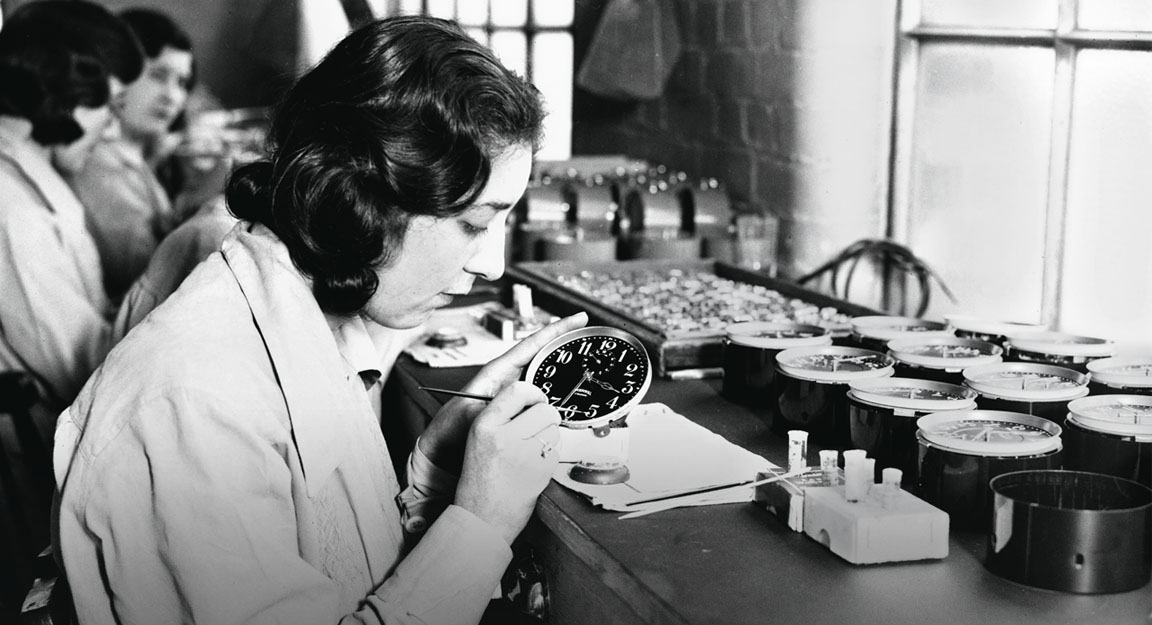
Female dial painter at the US Radium Corporation

What the Radium Girls Taught Us About Radiation Safety

The plight of the Radium Girls in the 1920s would teach us a great deal about the radioactive element radium and its effect on the human body. It brought to light the dangers of working with radium and created a universal understanding of the need for occupational and radiation safety measures.

What is radium?

Radium is a naturally occurring radioactive metal formed when uranium and thorium decay. In the environment, radium is present at low levels in groundwater, soil, rocks, and plants.

There are four radium isotopes, all of which are radioactive and have drastically different half-lives.

As radium decays it releases ionizing radiation in the form of alpha, beta, and gamma radiation. This radiation excites certain fluorescent chemicals in the metal and results in radioluminescence. It can also form other elements, such as radon.

Radium was discovered by Marie Sklodowska Curie and her husband Pierre Curie in 1898, although it would be more than a decade before the pair had isolated a sample large enough to work with.

Early Misinformation About Radium

Soon after the Curies discovered radium, medical professionals began using the radioactive substance as a cancer treatment. Before it could be properly studied, this initial use led to an explosion of interest from the American public and a host of false medical claims that radium was “healthful rather than medicinal.” 

Radium was initially considered a cure-all for a variety of health conditions, including arthritis, tuberculosis, rheumatism, gout, and high blood pressure. It was also thought to improve vitality in the elderly, treat skin conditions like eczema, and cure insomnia.

Because of its seemingly magical healing properties, major corporations began putting radium into their products and heavily promoting its use. Radium-infused toothpaste, pillows, facial creams, and tonic water were popular amongst the public, as were radium spas and clinics.

Radium-based cosmetics were trendy among women. They used these products to combat signs of aging in the form of wrinkles, crows-feet, and even unwanted body or facial hair.

The radium cosmetics gave the women’s skin a warm and cheerful glow and came to be known as “liquid sunshine.” This further cemented the idea that the products contained restorative properties that would revitalize the body and improve its overall health.

Who Were the Radium Girls?

In 1917, the United States entered World War I. There was a sudden demand for instruments and watches that could be read in the dark by U.S. soldiers. Thanks to a high-tech, glow-in-the-dark paint called UnDark, which was made with radium, this became possible.

With most of the country’s men on foreign battlefields, the United States Radium Corporation (USRC) in New Jersey began hiring young women to paint a variety of radium-lit instruments for use in the trenches. These women were called dial painters.

The dial painters would mix the radium-based paint in a crucible at their workstations and used fine, camel hair paint brushes to paint on the tiny, delicate numbers. The brushes quickly lost their shape after a couple of strokes. Management encouraged the women to use their lips to bring the brushes to a fine point for better precision. They were told repeatedly that radium was safe to ingest, and so continued with the “lip, dip, paint” process while they worked.

However, the dial painters didn’t just ingest the radium at their workstations. Due to its many reported health claims, workers would often paint their teeth or nails with radium-based paint before going out for the evening to impress their dates or amaze party guests. The dust from the hand-mixed paint coated the women’s hair and dresses, giving them a ghost-like glow that earned the women the nickname “ghost girls.”

For many dial painters, who were mostly between the ages of 14 and 20, this work was as desirable as it was glamorous. Radium’s luminous, sparkling appearance gave them a unique status. Furthermore, America’s obsession with its magical healing properties combined with the available compensation for the work had entire families flocking to the factory for a position.

Radiation Sickness

By the early 1920s, medical professionals throughout the area were noticing a frightening increase in the young worker’s health complaints. Many of their female patients complained of stiff and cracking joints, painful toothaches, oozing mouth sores, and listlessness, while others had broken out or developed severe anemia.

Dentists began pulling multiple teeth from young dial painters at a time. There were several instances where, during the tooth extraction, pieces of the woman’s decaying jawbone would come out with the tooth. In many cases, the tooth extractions wouldn’t heal.

Other symptoms of radium poisoning in the dial painters, which would later become understood as radiation sickness, were sterility, cataracts, leukopenia, eosinophilia, leukemia, anemia, and menstruation issues.

Mollie Maggia was the first dial painter to fall ill and die. She first developed increasingly painful toothaches that traveled from tooth to tooth. Severe pain in her limbs also prohibited her from walking.

Although dentists didn’t know it at the time, Mollie had developed “radium jaw.” This occupational disease involved necrosis of the upper and lower jawbones, bleeding gums, ulcers, and bone tumors. At the end of her life, Mollie’s dentists merely lifted her jaw from her mouth to remove it. Mollie died in 1922 just days before her 25th birthday.

Another 12 women who worked for the U.S. Radium Corporation as dial painters died the following year, with an additional 50 women falling severely ill.

Radium’s Effect on the Human Body

Radium has similar effects on the human body as calcium and strontium when inhaled or ingested. Once it enters the bloodstream, radium concentrates in the bones in high quantities. It emits alpha particles as it decays, which irradiates the cells on the bone’s surface. Over time radium will settle into the bone where it wreaks havoc on bone marrow and blood cell production.

If radium is ingested with food or water, over 80% of the element is excreted through urine or feces. The other 20% will travel throughout the body, settling in the bones and remaining there throughout the person’s life.

Historical Impact & Worker’s Rights

The surviving dial painters sued the U.S. Radium Corporation, although the road to doing so was not easy. The case was eventually settled out of court in 1928. The women were awarded $15,000 plus $600 per year for future medical expenses because of radium poisoning.

This landmark case was one of the first instances of workers receiving compensation for a disease developed because of their occupation. However, most of the women who received the money died within two years of the settlement.

At the time of the dial painters, there were no radiation safety measures put into place to prevent direct contact with the radioactive substance from occurring. The case of the Radium Girls opened people’s eyes to the dangers of radium and other radioactive substances. They were seen as an example of what could go wrong in an occupational setting and completely changed the course of occupational disease labor laws and regulations.

Their case had a direct impact on scientists’ approach to radiation safety during The Manhattan Project. It was also a leading cause for the creation of the Occupational Safety and Health Administration in 1970.

Radium would continue to be used as a luminescent paint until the early 1960s when its toxicity and danger to human life could no longer be ignored.

29 Jun 2022
Straw hat, bright red glasses and orange bottle of sunscreen for sun protection

Ultraviolet Radiation: How to Protect Yourself

Summer is in full swing. As a result, many Michiganders are spending more time in the great outdoors taking advantage of the warmth and sunshine.

However, the more time a person spends outdoors the more their body is exposed to Ultraviolet (UV) radiation. UV radiation, a form of non-ionizing radiation, is invisible to the human eye and cannot be felt. It can cause severe skin damage and lead to the development of skin cancer.

Here at Versant Physics, our focus is primarily on radiation safety in relation to ionizing radiation sources used in medical procedures and cancer treatments. However, it is just as important that you protect yourself from naturally occurring UV radiation and understand the potential health risks.  

In honor of UV Safety Month, we’ll explain what UV radiation is, the most common types of skin cancer and other health risks associated with UV radiation, and important protective measures you should take when out in the sun.

What is UV Radiation?

Ultraviolet (UV) radiation is a non-ionizing form of electromagnetic radiation that has both natural and artificial sources. Most of the UV radiation from sunlight gets absorbed by Earth’s atmosphere. What doesn’t get absorbed makes its way to the surface and interacts with our skin. UV rays are present even on cloudy days and also reflect off surfaces like snow, sand, and water.

There are three types of UV radiation rays:

  • Ultraviolet A (UVA)
  • Ultraviolet B (UVB)
  • Ultraviolet C (UVC)

UVA rays have the lowest wavelength of the different types of UV radiation; however, they make up over 95% of the rays that reach the Earth’s surface. These rays penetrate through the layers of skin, damaging the elastin and collagen. This results in tanned skin and skin aging, often in the form of wrinkles or age spots.  

UVB radiation is made up of high-energy UV rays that interact with the top layers of skin. UVB rays interact with skin cells and damage them, causing DNA mutations that show up later in the form of sunburns, skin cancer, or cataracts.

UVC rays are the strongest of the UV rays. Almost all of this UV radiation is absorbed by Earth’s atmosphere.

Unprotected, prolonged exposure to UV radiation from the sun is connected to a variety of health risks, including:

  • Premature aging
  • Skin damage
  • Cataracts
  • Immune system suppression
  • Skin cancer

UV Radiation Exposure and Skin Cancer

UV radiation causes melanoma and nonmelanoma skin cancers called basal cell carcinoma (BCC) and squamous cell carcinoma (SCC).

Melanoma

Melanoma is a type of skin cancer that forms in the melanocytes. These cells are located beneath the squamous and basal cells and are what produce melanin, the pigment that gives hair, eyes, and skin its color.   

Melanoma is less common than other types of skin cancer, however, it is more dangerous. This is because melanoma it more likely to spread to other parts of the body if left untreated. Melanoma often presents as a highly pigmented black or brown tumor on the torso, chest, neck, or face.

The “ABCDE” rule can help patients identify if their existing mole or new skin growth is a warning sign of melanoma:

  • Asymmetry. The two halves of the mole do not match.
  • Border. Normal moles have a clean, even border. Melanoma will present with uneven or ragged edges.
  • Color. Melanoma tumors can be black, brown, pink, red, or white.
  • Diameter. Melanoma is usually a growth larger than a pencil eraser, or ¼ inch in diameter.
  • Evolving. The existing mole or new growth is changing in size, shape, texture, or color. It also may begin to itch, bleed, or ooze.

People with light skin, eyes, and hair are considered more at risk of developing melanoma than people with darker skin. Age, gender, occupation, family history, and lifestyle choices also play a role in the level of risk associated with developing melanoma.

Nonmelanoma Skin Cancers

Basal cell carcinoma is the most common type of skin cancer that begins in the basal cells. It shows up on areas of the body that are frequently exposed to UV radiation from sunlight, such as the head, face, or neck. It normally presents in the form of a skin lesion or shiny, skin-colored bump.

Squamous cell carcinoma is less common but just as serious. It presents as open sores, thick or wart-like skin, raised growths, or scaly red patches that may itch or bleed. SCC can show up anywhere on the body, although they are most often found on areas of the body that are frequently exposed to the sun.

Both BCC and SCC grow relatively slowly and are highly treatable. The sooner a new or strange-looking growth is looked at by a dermatologist and diagnosed, the better the odds are of treating the skin cancer. However, if left untreated, these skin cancers can spread to other areas of the body and become more dangerous.

UV Radiation Protection

There are many simple protective measures the average person can implement to help lessen the risks associated with UV radiation and its negative side effects.

  • Seek shade
  • Avoid prolonged sun exposure from 10 a.m. to 4 p.m. when the sun is strongest
  • Wear long sleeves or pants
  • Wear a hat and/or UV-blocking sunglasses
  • Wear a broad-spectrum sunscreen

Sunscreen is a major protector against UV radiation. Broad-spectrum sunscreens protect the skin from both UVA and UVB rays. Wearing a minimum of SPF 15 can reduce the risk of developing melanoma by 50% and SCC by 40%. Wearing a protective sunscreen daily can also help prevent premature skin aging.

Are There Any Health Benefits to UV Radiation?

Exposure to natural UV radiation from the sun has an important health benefit for the human body. UV radiation helps our bodies produce vitamin D, which is an essential vitamin that absorbs calcium in our stomachs, reduces inflammation, and is needed for healthy bone growth. Some food products contain vitamin D however most people get a portion of their vitamin D needs through sunlight.

There are no hard and fast numbers detailing how much sunlight exposure is needed for optimal vitamin D synthesis. The World Health Organization recommends no more than 15 minutes of direct sun exposure at least 3 times a week.

However, this does not negate the need for sun protection measures such as sunscreen and wearing protective clothing.

The Takeaway

It is important to protect yourself from UV radiation any time of the year. Although this non-ionizing source of radiation can help our bodies create vitamin D, it also interacts with our skin in a way that can lead to skin cancer. To prevent this, you should wear and apply sunscreen as directed, invest in UV-blocking glasses and clothes, and try to stay out of the sun as much as possible.

Learn more about UV and sun safety here.

20 Apr 2022
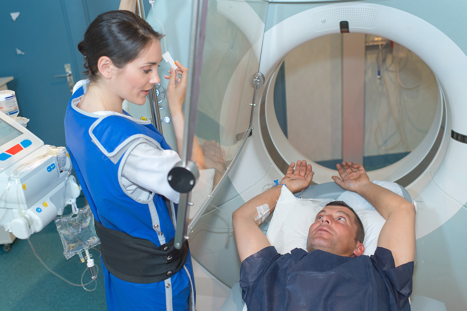
nurse guiding patient entering mri scanner

The Basics of Radiation Shielding in Medicine

Basic radiation protection guidelines can be summed up in three simple concepts: time, distance, and shielding. While both limiting the time spent and increasing the proximity to an ionizing radiation source is something that lies within the power of the individual, shielding and X-ray room design require careful planning and execution by the facility or Radiation Safety Officer.

What is radiation shielding?

Radiation shielding is simply a barrier placed between a source of radiation and the area or person that needs to be protected. The purpose of radiation shielding is to limit, control, or modify the radiation exposure rate at a set point.

Shielding is based on attenuation or the gradual reduction in the intensity of energy through a specific medium. X-ray radiation that passes through certain materials decrease and are absorbed, thereby reducing the exposure to the other side of the barrier.

Without shielding, the public, radiation workers (including dentists and veterinarians), and even nearby office workers could be exposed to levels of radiation outside regulated exposure limits, which can potentially lead to negative health effects. Although it is impossible to completely avoid exposure to radiation, shielding is a critical consideration in any medical facility that greatly reduces unnecessary exposure.

Shielding Materials

There are several different materials that provide protection from penetrating radiation. Concrete, water, special plastic shields, air stops, and lead are all barriers that stop different types of rays and particles, reducing the overall dose a person receives.

In medical environments, the most common shielding materials used include lead, lead-free shielding, and lead composites.

Lead

Lead is one of the most used and most effective shielding materials. It is a highly dense material with a high atomic number and a high number of electrons which make it ideal for shielding in most medical radiation environments. This is because the type and energy of radiation in a medical environment that passes through lead are absorbed or scattered by the electrons present in the material.

Vet team wearing shielding garments during exam

Lead is also cheap and easy to process. It can be mixed with other materials like glass, or binders like vinyl, which allows it to be used as construction materials in X-ray rooms or worn as shielding garments.

Lead-Free Shielding

Technological advances have allowed for the creation of non-toxic, lead-free shielding materials as well. Other attenuating materials such as antimony (Sb), tungsten (W), and tin (Sn) are used in place of lead and combined with additives and binders to create wearable protective garments or materials. They offer equal protection from scatter radiation.

Lead-free shielding has several benefits, including being both recyclable and non-toxic. Lead-free shielding materials can also be lighter which makes them easier for personnel to wear during longer procedures.  

Lead Composite

Lead composite shielding is a long-lasting mixture of lead and lighter materials that attenuate radiation just as successfully as traditional lead shielding barriers.

Because of lead’s weight, it can be cumbersome to use and wear for long periods of time, limiting the efficacy of a radiation worker. Lead composites solve this problem. They are made with blends of tin, vinyl, and rubber and create a shielding barrier that can be up to 25% lighter than traditional shielding without sacrificing their ability to block penetrating radiation.

Shielding and Scatter Radiation

In some diagnostic X-ray procedures, medical personnel such as operators, radiologists, and technologists are required to remain in the room with the patient. This proximity frequently exposes them to something called scatter radiation or radiation that bounces off a patient’s body during a procedure.

To limit this exposure, some medical personnel are required to wear frontal or full wrap-around style lead aprons, thyroid shields, and lead glasses/gloves. These protective garments can attenuate roughly 93% of photons at typically scattered energies.

Lead apron and thyroid collar on hangar

Shielding Products and Design

There are several different ways radiation shielding can be applied or designed to protect healthcare workers.

Room Shielding

Shielding may be required in the floor, ceiling, doors, or any wall of any X-ray or radioactive material use room.  Shielding is used to protect workers, patients, or the public that may be in the adjacent areas/rooms.

During a room’s construction, special shielding materials are installed where their need has been determined. These materials can include lead-lined windows and doors, lead-lined drywall or plywood, lead sheets for floors and ceilings, pipe shielding, and more.

X-ray room shielding requirements vary from state to state. It is important to consult with a qualified expert familiar with these regulations as well as work with an architect experienced in constructing X-ray suites before building a new room.

Leaded Glass and Curtains

In some cases, it isn’t possible for a facility to build shielding into the physical structure of a building.

Leaded glass barriers are a barrier used by techs and doctors which allow them to safely view a patient during an imaging procedure. This type of glass is ideal for radiation-producing equipment in the 80-300 kV range thanks to its high lead content.

Lead curtains are also used to shield radiation workers, particularly in large animal hospitals or operating rooms. These curtains are leaded rubber or vinyl sheets that are ideal for protection against low-level or secondary radiation. They make for room-saving partitions that can be open or closed as needed and typically offer protection from 0.5mm to 2.00mm lead equivalency.

Mobile Shielding Barriers

In some cases, additional barriers are needed to protect doctors and techs during radiology, nuclear medicine, cath lab, or diagnostic imaging procedure. These barriers are lead-lined partitions on wheels, often with a protected window to allow for patient observation.

Mobile radiation barriers come in a variety of shapes, sizes, and lead equivalencies. They are ideal for maintaining flexibility and ease of movement in a procedure room while successfully minimizing the scattered radiation dose to workers in the room.

Versant Physics Shielding Services

Understanding the detailed shielding requirements for your state or facility can be a time-consuming challenge. If executed incorrectly, there can be serious consequences to the health and safety of radiation workers, patients, and building staff as well as potential regulatory compliance fines.

That’s why it is important to have a radiation safety consultant like Versant Physics on your side. Whether you’re constructing a new X-ray room, remodeling or repairing an existing shielding setup, or looking to upgrade your current shielding equipment, our team of expert health and medical physicists can assist.

We provide radiation shielding calculations, evaluation, and design for facilities of all kinds, including hospitals, clinics, dentist offices, chiropractor offices, and veterinary clinics. Our range of expertise includes:

  • Radiography
  • Fluoroscopy
  • Computed Tomography (CT)
  • Nuclear Medicine/PET
  • Mammography
  • Dental/Veterinary X-ray

Not sure what materials or type of shielding is right for your facility? Contact our regulatory experts for a free 30-minute consultation.

05 Jan 2022

Top 3 Consumer Products that Contain Radioactive Materials

Radioactive materials are present in our natural environment and in man-made products we use every day. Such consumer products are defined as “a device or manufactured item into which radionuclides have deliberately been incorporated or produced by activation, or which generates ionizing radiation, and which can be sold or made available to members of the public without special surveillance or regulatory control after sale.”

Many devices that use WiFi or Bluetooth technology or connect to cell phone towers emit radio waves, also known as electromagnetic radiation (EMF).

This may concern consumers who are worried about the negative health effects associated with “radioactive materials” and “radiation.” However, in most cases, these materials we interact with are safe and pose no danger to our health.

Below we guide you through three common consumer products the average person uses or engages with regularly, discuss how the radioactive materials they contain work, and determine the health risk they pose to you and your family.

Cell Phones

Cell phones have become an integral part of daily modern life. We depend on them for communication, connection, and as a source of entertainment. However, their permanent presence and increased usage have raised concerns over the years that cell phones can cause negative health effects to humans, including brain tumors and hearing loss.

pile of cell phones

Do cell phones emit radiation?

Cell phones are not consumer products that contain radioactive materials. However, they communicate by transmitting EMF, a type of non-ionizing radiation at the low-energy end of the electromagnetic spectrum in the 100kHz to 300GHz frequency range.

RFs are widely used in communication technologies such as cell phones, Wi-Fi, radio, and TV. They are also found in MRI equipment, from natural sources like outer space, and in the microwave oven sitting on your kitchen counter.

Are there health risks?

Decades of research on RF radiation have concluded that exposure to this frequency has minimal health effects. Due to their frequency, RF radiation can be absorbed by the human body. In large amounts, this can produce heat, which has the potential to cause burns or tissue damage.

Numerous short-term studies have taken place on the link between cancer rates and cell phone usage. Small, individual studies have found slight associations between cell phones and cancer of the salivary glands, as well as a possible increase in the risk of gliomas. In 2011, the International Agency for Research on Cancer evaluated these studies and concluded that there is limited or inadequate evidence of carcinogenicity. Longer-term studies may need to be conducted to accurately determine the level of cancer risk associated with cell phones.

Those uncomfortable with incurring any level of risk can take steps to limit their cell phone usage by purchasing a hands-free headset or utilizing the speakerphone function when making calls.

Smoke Detectors

Most smoke detectors in the United States are ionization smoke alarms, which contain a small amount of the man-made radioactive element called americium-241.

how smoke alarms work

Why is radioactive material present?

Ionization smoke alarms are more responsive to flaming fires. The radioactive material present in the smoke alarm rests between two electrically charged plates which ionize the air and causes a current between them. Smoke entering the chamber disrupts the flow of ions, reducing the current and thereby activating the alarm.

Are there health risks?

Smoke detectors pose little to no health risk to human beings. The amount of americium-241 present is minimal, wrapped in gold foil, and shielded by the plastic case and stainless steel. These protective measures prevent easy tampering rather than limiting radiation exposure. However, there is no risk of significant exposure as long as these sources are contained in the detector housing.

Granite Countertops

Like many natural materials found on Earth, granite, a type of durable stone used in construction and home décor, contains small amounts of radioactivity.

Granite is a consumer product that contains a small amount of natural radioactive material.

Does granite emit radiation?

Trace elements of uranium, thorium, and radium can show up in slabs of granite. When these elements are present, they decay into radon. According to the EPA, radon released from granite materials can be released over the lifetime of its use but is typically diluted by ventilation.

Are there health risks?

It is extremely unlikely that the radiation emitted from granite countertops in your home would increase radiation doses above normal background levels. The radon released from granite is a significantly lower concern when compared with radon which originates in the soil and can build up inside the home. This type of radon is the second leading cause of lung cancer in the United States and should be tested for on a regular basis.

Conclusion: Are Consumer Products That Contain Radioactive Materials or Emit Radition Unsafe?

It is true that some common consumer products contain trace amounts of naturally occurring radioactive materials or emit non-ionizing radiation. However, this does not mean they are dangerous or pose a health risk to humans. In fact, in products like ionizing smoke detectors, the presence of radioactive material is crucial for keeping humans safe.

Further Reading:

Radiation Safety for Consumer Products, Specific Safety Guide No. SSG-36

15 Jun 2021

The Truth About Background Radiation

Background radiation is all around us, and always has been. That idea can be a frightening concept at face value, but the truth is background radiation is natural, normal, and expected.

Most natural background sources of radiation fall into one of three categories:

Cosmic Radiation

Think of this as steady waves of external radiation being sent from the sun and stars in space to Earth. This type of radiation occurs naturally and introduces extremely low levels of radiation to the average person. The amount (or dose) of cosmic radiation one receives can depend on weather and atmospheric conditions, the Earth’s magnetic field, and differences in elevation. For example, people who live at higher altitudes like Denver, Colorado are exposed to slightly more cosmic radiation than people who live in lower altitudes, such as New Orleans, Louisiana or Miami, Florida. Furthermore, the farther north or south one is from the equator results in a higher dose of cosmic radiation due to the way the Earth’s magnetic field deflects cosmic radiation toward the North and South poles.

silver airplane flying above orange clouds

Air travel can also expose individuals to low levels of cosmic radiation. The received dose is similarly dependent on altitude, latitude, and the duration of the flight. A coast-to-coast flight in the United States would expose an individual to approximately 3.5 mrem. For comparison, a typical medical procedure involving radiation, such as a chest X-ray, exposes an individual to 10 mrem, and the average American receives a total radiation dose of 540 mrem each year.

In general, a person’s average dose from cosmic radiation in the United States is small, making up only 6% of their total annual dose.

Terrestrial Radiation

Terrestrial radiation is the portion of natural background radiation that is emitted by naturally occurring radioactive materials on earth, and it is responsible for approximately 3% of the average person’s annual received dose. The physical earth, including soil and sedimentary and igneous rock, contains common elements like uranium, thorium, and radium. These naturally occurring radioactive materials, which have existed as part of the earth’s crust since the earth was formed, are released into the water, vegetation, and the atmosphere as they breakdown at different rates. People are largely exposed to the resulting emitted radiation through their skin.

Radon:

diagram of radon gas infiltrating a house

Perhaps the most significant form of terrestrial radiation is that which is inhaled. When the naturally occurring radioactive element uranium (found in the earth’s crust, underwater caves, and seawater) decays it can change into a scentless, invisible gas called radon. All the air we breathe contains trace amounts of radon, and it is responsible for the largest portion of background radiation dose that the average American receives in a year. Outdoors, this radioactive gas disperses rapidly and does not pose any health risk to human beings. A build-up of radon gas indoors, however, can potentially increase the risk of lung cancer over time, which is why it is important to test homes and workplaces for radon on a regular basis. Smoking, especially near or inside the home, can amplify the risk of cancer when coupled with radon exposure.

The average person can expect to receive 42% of their annual radiation dose from radon.

Internal Radiation

Background radiation can also be received through ingestion. Some common foods contain small amounts of radioactive elements that do not pose a radiation risk to the person ingesting them. The most common example is the banana. This delicious, nutritious fruit contains naturally high levels of potassium which helps muscles contract, keeps your heartbeat regular, and offsets the harmful effects of sodium on blood pressure. A tiny portion of potassium is also naturally radioactive. A single banana emits 0.01 mrem, which is received internally by the person eating it. According to the EPA, a person would have to eat 100 bananas to receive the same amount of radiation exposure naturally received each day from the environment. (It should be noted that this naturally occurring radiation is not the same thing as food irradiation, which is a process used by humans to kill bacteria, molds, and pests to prevent foodborne illnesses and spoilage.) Overall, the levels of natural radionuclides found in our food and water are low and considered safe for human consumption by regulatory bodies.

Most surprisingly for some is the fact that other humans are also a source of exposure to one another. From birth, people have internal radiation in the form of radioactive potassium-40, lead-210, and carbon-14. These elements reside in our blood and bones. As previously noted, humans also ingest traces of naturally occurring radioactive material found in our food and water. When our bodies metabolize the non-radioactive and radioactive forms of potassium and other elements, they then contain small amounts of radiation which can act as exposures to others.

Man-Made Radiation Exposure

A more familiar source of radiation exposure to many is man-made radiation, such as procedures using X-Rays and radiation therapy to treat cancer. According to the Health Physics Society, approximately 42% of annual dose comes from man-made radiation. This percentage includes medical procedures, household products like smoke detectors, and small quantities of normal discharges from nuclear and coal power plants.

Learn more about the health effects of man-made ionizing radiation in our blog post here.

Conclusion

Natural background radiation has always been a part of life on earth, and it always will be. It is important to understand that this is not something to be feared. Low levels of ionizing radiation from naturally occurring sources such as space, the ground beneath our feet, and even some of the food we eat are not dangerous and do not pose a direct health risk to ourselves or our loved ones.

For more information, visit the Health Physics Society webpage, epa.gov, or the International Atomic Energy Agency.

Note: Visit our regulatory page to learn how Versant Physics’ board-certified Internal Dose Specialists, Medical Physicists, and Health Physicists, can assist with your radiation safety program needs.

Additional Sources:

https://www.nrc.gov/about-nrc/radiation/around-us/sources/nat-bg-sources.html

https://www.cdc.gov/nceh/radiation/air_travel.html

NCRP Report 160

NCRP Report 184

20 May 2021
Smiling pregnant worker

Occupational Radiation Workers & Declaring a Pregnancy

Employees who become pregnant and work with radiation or radioactive materials during their pregnancy are often concerned about the safety of doing so, as well as the potential effects of radiation on their unborn child. Occupationally exposed workers are not required to declare a pregnancy to their employer. However, if they decide to declare there are dose limits that should be observed and additional protective measures that can be taken to protect both mom and baby.

Declaring a Pregnancy


In the United States, pregnant employees who work with or around radiation have the option of declaring their pregnancy. This declaration is voluntary and informs the worker’s employer in writing of their pregnancy as well as the estimated date of conception. This information is confidential and shared only with the employer and radiation safety officer, however, it is valuable for reducing exposure and allowing for close monitoring of both the employee and the baby throughout the pregnancy.

pregnant radiation worker consulting with radiation safety officer

The NRC and States require licensees and registrants (i.e., the facility the employee works at) to make efforts to limit the declared pregnant worker’s received dose. This can mean that some normal job functions may not be permitted if doing those jobs would result in the fetus/embryo receiving more than 500 mrem.

Employees also have the option to discuss with their employer or radiation safety officer about potential changes to their job status prior to declaring a pregnancy if they so choose. The option to revoke a declaration of pregnancy even if the worker is still pregnant is also available at any time throughout the pregnancy.

When an employee declares a pregnancy, they should sit down with their radiation safety officer for a one-on-one counseling session. This is a great opportunity to ask questions and address any monitoring or safety concerns that may arise.

They are then issued a fetal dosimeter in addition to their regular monitoring device, which is worn at the hip or waist level. For procedures where a lead apron is worn, the dosimeter should be worn beneath it while the regular dosimeter is worn on the outside at the neck or collar. The fetal dosimeter is monitored monthly by the radiation safety officer to ensure that the regulatory fetal dose limits are not exceeded.

According to regulations, the lower dose limit for the embryo or fetus remains in effect until the worker withdraws the declaration in writing or is no longer pregnant. If it is not withdrawn, the original declaration expires after one year.

If an employee chooses not to declare their pregnancy, the employee and her baby are restricted to the standard occupational dose limits that apply to all occupationally exposed workers. The annual total effective dose equivalent (TEDE) for the whole body is 5,000 mrem. (10 CFR Part 20.)

Occupational Exposure


In most cases, the ways in which a pregnant woman may be occupationally exposed to radiation within regulatory limits are not likely to cause adverse health effects for the developing fetus.  However, most regulations are guided by the principle that any level of radiation can potentially result in negative biological effects and that the likelihood of such effects increases as the dose received increases.

The NRC requires licensees to “limit exposure to the embryo/fetus of an occupationally exposed individual to 500 mrem (5 mSv) or less during pregnancy for a declared pregnant worker who is exposed to radiation from licensed radioactive materials including radionuclides.” (10 CFR 20.1208) This lower dose limit is “based on a consideration of greater sensitivity to radiation of the embryo/fetus and the involuntary nature of the exposure.”  

Pregnant nurse on the phone with ipad

To break this down further, the regulations state that the radiation dose from occupational exposure should be limited to 500 mrem for the duration of the pregnancy and no more than 50 mrem per month. At this level, (1/10 the dose that a regular occupationally exposed worker may safely receive in a year) the risk of negative health effects is low.

Pregnant workers can speak directly with their radiation safety officer or on-site medical or health physicist to determine the safest dose limits for their individual needs, which may depend on their exposure history and the types of jobs they perform on a regular basis.

Undergoing Medical Procedures While Pregnant


Occupational limits for declared pregnant workers do not apply to individuals who undergo diagnostic or therapeutic procedures, such as X-rays, fluoroscopy, or radiation therapy.

According to Robert Brent, MD, Ph.D. for HPS.org, diagnostic procedures of different parts of the body, such as the head, teeth, legs, or arms do not directly expose the fetus. Modern medical imaging procedures focus the X-ray beam only on the body part of interest, and the amount of radiation that could reach the embryo or fetus during these diagnostic procedures is small and unlikely to increase the risk of miscarriage or birth defects. Most procedures expose the developing fetus or embryo to less than 50 mSv, if at all. At this level of exposure, there is no cause for concern.

Regardless of pregnancy status, the ALARA principle should be implemented by the individual’s care team to guide decisions made about treatment and diagnostic procedures. A radiation safety officer or medical physicist can also help provide options to minimize dose. It should also be noted that those with fetal dosimeters should not wear their dosimeter during an X-ray or nuclear medicine procedure.

Conclusion


Ultimately, the decision to declare a pregnancy is that of the pregnant radiation worker. Under the current safety guidelines, the risk for adverse health effects to an embryo or fetus posed by occupational exposure or medical procedures is low. However, employees should take advantage of the resources available such as the NRC regulations, literature provided by the Health Physics Society, and the expertise of their radiation safety officer and on-site medical or health physicist.

Visit our website to learn more about Versant Physics regulatory services, including radiation safety officer support, personnel dosimetry management for declared pregnant workers, and more.


Sources

https://www.cdc.gov/nceh/radiation/emergencies/prenatalphysician.htm

https://www.nrc.gov/reading-rm/doc-collections/cfr/part020/index.html

https://hps.org/hpspublications/articles/pregnancyandradiationexposureinfosheet.html

https://www.ncbi.nlm.nih.gov/pmc/articles/PMC3835582/