Category: Ionizing Radiation

27 Mar 2024
Airplane flight crew character design. Pilot and stewardess flat vector illustration

Flight Crews and Radiation Exposure

Flight crews are among the occupational groups most exposed to ionizing radiation, with an average annual effective dose surpassing that of other radiation-exposed workers in the United States, excluding astronauts.1 This elevated exposure is primarily due to the high levels of cosmic radiation encountered at flight altitudes, which can pose significant health risks to pilots and cabin crew members.2 In this blog post, we’ll explore the nature of cosmic radiation, its potential health effects, current exposure levels for aircrews, as well as the guidelines and regulations in place to ensure their safety.

What Is Cosmic Ionizing Radiation?

As we’ve touched on in a previous blog, cosmic ionizing radiation–or simply cosmic radiation–originates from beyond Earth’s atmosphere. Additionally, it consists of two main components: galactic cosmic radiation (GCR) and solar particle events (SPEs).3,4

Galactic Cosmic Radiation

GCR is a constant background radiation that permeates interstellar space, originating from distant stars and galaxies. It is composed primarily of high-energy protons (85%) and alpha particles (14%). There is also a small fraction of heavier nuclei (1%) ranging from lithium to iron and beyond. These particles span a wide energy range, and as a result some reach extremely high energies capable of penetrating deep into the Earth’s atmosphere and passing through aircraft shielding.5

Solar Particle Events (Solar Flares)

Solar particle events, on the other hand, are sporadic bursts of intense radiation associated with solar flares and coronal mass ejections. During an SPE, the Sun ejects a large number of high-energy protons and other particles that can reach Earth within hours to days. While less frequent than GCR, SPEs can dramatically increase radiation exposure for flight crews, particularly those on polar routes where the Earth’s magnetic field provides less protection.6,7

At higher altitudes, such as those typically encountered during air travel, the Earth’s atmosphere provides less shielding against cosmic radiation, resulting in increased exposure for flight crews and passengers.

Several studies that have investigated the difference in cosmic ray levels at various altitudes versus ground level found that the dose rate of cosmic radiation at a cruising altitude of 30,000 feet was approximately 10 times higher than at sea level.

The specific increase in cosmic ray exposure at higher altitudes is influenced by several factors, including the solar cycle (solar maximum vs. solar minimum), geomagnetic field strength, and also the path of the flight (polar routes are exposed to higher levels of cosmic rays). For example, during periods of high solar activity (solar maximum), the increased solar wind can actually shield the Earth from some cosmic rays, slightly reducing the exposure at high altitudes. Conversely, during a solar minimum, the cosmic ray intensity can be higher.

Estimates of the number of hours that have to be flown in order to receive an effective dose of 1 mSv at 30,000 feet are 510 hours at a latitude of 30o South and 1,330 hours at the equator.8

Health Effects and Uncertainties

The health risks associated with radiation exposure are generally well-documented. Prolonged exposure to high levels of radiation can increase the risk of cancer, cataracts, as well as other adverse health effects.  However, quantifying the specific risks associated with the chronic low-dose radiation experienced by flight crews remains a challenge.

The World Health Organization’s International Agency for Research on Cancer (IARC) acknowledges that ionizing radiation causes cancer in humans and is also associated with reproductive problems. However, when it comes to cosmic ionizing radiation, several uncertainties remain:

  1. Cancer Risk: Most radiation health studies have focused on groups exposed to much higher doses from different types of radiation (such as atomic bomb survivors or patients receiving radiation therapy).9 Due to this, the specific link between cosmic ionizing radiation and cancer risk is not yet fully understood.
  2. Reproductive Health: Miscarriages and birth defects related to cosmic radiation exposure are still not definitively established.10

Despite the limitations of current research, several studies have suggested that flight crews may have a higher incidence of certain cancers compared to the general population. These include breast cancer, melanoma, as well as non-melanoma skin cancers.11,12 However, the causal link between cosmic radiation exposure and these increased risks has not been definitively established. Other factors, such as lifestyle and genetic predisposition, may also play a role.13

Exposure Levels for Flight Crews

Recent dose and risk assessments by a wide variety of investigators have demonstrated the need to dedicate further attempts to quantify potential radiation exposure.14 The National Council on Radiation Protection and Measurements (NCRP) reports an average annual effective dose of 3.07 mSv for flight crews; most of this exposure comes from natural radiation:

  • Estimates of annual aircrew cosmic radiation exposure range from 0.2 to 5 millisieverts (mSv) per year depending on factors such as flight routes, altitude, and solar activity.
  • Solar particle events occur less frequently, but during events, exposure levels can increase substantially and potentially lead to higher doses over short periods.

Guidelines and Regulations

While there are no official dose limits specifically for aircrew in the United States, national and international guidelines provide context:

  • International Commission on Radiological Protection (ICRP): Recognizes aircrew as radiation-exposed workers. They also recommend an effective dose limit of 20 mSv per year averaged over 5 years (totaling 100 mSv in 5 years) for radiation workers. However, for the general public, the recommended limit is 1 mSv per year.15
  • Pregnant Aircrew: The ICRP recommends a dose limit of 1 mSv throughout pregnancy.16

Current regulations aim to limit radiation exposure for flight crews, but there is room for improvement. The International Commission on Radiological Protection (ICRP) sets guidelines for radiation protection and also includes dose limits for occupational exposure. However, these guidelines may not adequately address the unique challenges faced by flight crews. To improve current radiation safety regulations for aircrews, a multi-faceted approach is necessary. This should include:

Improved Monitoring and Data Collection

Implementing advanced radiation monitoring systems on aircraft in addition to encouraging the use of personal dosimeters by flight crews can provide more accurate and comprehensive data on exposure levels17. This information can help refine risk assessments as well as guide the development of more effective protection strategies.

Aircraft Shielding and Design

Continued research into advanced shielding materials in addition to aircraft design modifications can help reduce the radiation dose received by flight crews and passengers18. This may also involve the use of novel composite materials or the incorporation of additional shielding in critical areas of the aircraft.

Route Optimization and Flight Planning

By carefully planning flight routes and altitudes, airlines can minimize exposure to cosmic radiation, particularly during solar particle events19. This may also involve rerouting flights to lower latitudes or reducing flight time at higher altitudes when necessary.

Education and Awareness Programs

Providing flight crews with comprehensive information about the risks of cosmic radiation exposure in addition to the importance of proper protection measures can empower them to make informed decisions about their health and safety20. This should include training on the use of personal protective equipment, such as dosimeters, as well as guidelines for managing exposure during pregnancy.

Regulatory Harmonization and Enforcement

Strengthening international collaboration to harmonize radiation protection standards for flight crews in addition to ensuring consistent implementation and enforcement of these standards across the aviation industry can help create a safer working environment for all aircrews2121.

Conclusion

Although no regulations officially set dose limits, radiation exposure is still a concern to be evaluated for airplane flight crews due to their occupational exposure to cosmic radiation. While the specific health risks associated with this chronic low-dose exposure remain uncertain, continued efforts are essential ensure a safe working environment. By implementing measures such as personal dosimetry devices, increased monitoring, staff training, and encouraging airplane manufacturers to consider shielding and design modifications, airlines can better protect their flight crews. Ensuring a safer career for every radiation worker will require time, dedication, and collaboration. However, the benefits for the health and safety of all industries, including aircrews, make it a worthwhile endeavor.

Versant Physics is a full-service medical physics and radiation safety consulting company based in Kalamazoo, MI. Contact us for all of your regulatory, radiation safety, and personnel dosimetry needs.

Sources

  1. Friedberg, W., & Copeland, K. (2003). What aircrews should know about their occupational exposure to ionizing radiation. Oklahoma City, OK: Civil Aerospace Medical Institute, Federal Aviation Administration. ↩︎
  2. United Nations Scientific Committee on the Effects of Atomic Radiation. (2008). Sources and effects of ionizing radiation: UNSCEAR 2008 report to the General Assembly, with scientific annexes. New York: United Nations. ↩︎
  3. Validation of modelling the radiation exposure due to solar particle events at aircraft altitudes. Radiation Protection Dosimetry, Volume 131, Issue 1, August 2008, Pages 51–58. https://doi.org/10.1093/rpd/ncn238 ↩︎
  4. Wilson, J. W., Townsend, L. W., Schimmerling, W., Khandelwal, G. S., Khan, F., Nealy, J. E.,  & Norbury, J. W. (1991). Transport methods and interactions for space radiations. NASA Reference Publication, 1257 ↩︎
  5. O’Sullivan, D. Exposure to galactic cosmic radiation and solar energetic particles. Radiat Prot Dosimetry. 2007;125(1-4):407-11. https://pubmed.ncbi.nlm.nih.gov/17846031/ ↩︎
  6. Turner, R. E. (2007). Solar particle events from a risk management perspective. Radiation Protection Dosimetry, 127(1-4), 534-538. ↩︎
  7. Lantos, P., & Fuller, N. (2003). History of the solar particle event radiation doses on-board aeroplanes using a semi-empirical model and Concorde measurements. Radiation Protection Dosimetry, 104(3), 199-210. ↩︎
  8. Cosmic Radiation Exposure for Casual Flyers and Aircrew, https://www.arpansa.gov.au/understanding-radiation/radiation-sources/more-radiation-sources/flying-and-health ↩︎
  9. National Research Council. (2006). Health risks from exposure to low levels of ionizing radiation: BEIR VII phase 2 (Vol. 7). National Academies Press. ↩︎
  10. CDC – Aircrew Safety and Health – Cosmic Ionizing Radiation – NIOSH Workplace Safety & Health Topics. Centers for Disease Control and Prevention. Published 2019. https://www.cdc.gov/niosh/topics/aircrew/cosmicionizingradiation.html ↩︎
  11. Pukkala, E., Aspholm, R., Auvinen, A., Eliasch, H., Gundestrup, M., Haldorsen, T., & Tveten, U. (2003). Cancer incidence among 10,211 airline pilots: a Nordic study. Aviation, Space, and Environmental Medicine, 74(7), 699-706. ↩︎
  12. Rafnsson, V., Hrafnkelsson, J., & Tulinius, H. (2000). Incidence of cancer among commercial airline pilots. Occupational and Environmental Medicine, 57(3), 175-179. ↩︎
  13. Hammer, G. P., Blettner, M., & Zeeb, H. (2009). Epidemiological studies of cancer in aircrew. Radiation Protection Dosimetry, 136(4), 232-239. ↩︎
  14. Olumuyiwa A. Occupational Radiation Exposures in Aviation: Air Traffic Safety Systems Considerations. International Journal of Aviation, Aeronautics, and Aerospace. Published online 2020. doi:https://doi.org/10.15394/ijaaa.2020.1476 ↩︎
  15. International Commission on Radiological Protection. (2007). The 2007 recommendations of the International Commission on Radiological Protection. Annals of the ICRP, 37(2-4), 1-332. ↩︎
  16. International Commission on Radiological Protection. (2000). Pregnancy and medical radiation. Annals of the ICRP, 30(1), iii-viii, 1-43. ↩︎
  17. Bartlett, D. T. (2004). Radiation protection aspects of the cosmic radiation exposure of aircraft crew. Radiation Protection Dosimetry, 109(4), 349-355. ↩︎
  18. Wilson, J. W., Miller, J., Konradi, A., & Cucinotta, F. A. (1997). Shielding strategies for human space exploration. NASA Conference Publication, 3360. ↩︎
  19. Copeland, K. (2014). Cosmic radiation and commercial air travel. Radiation Protection Dosimetry, 162(3), 351-357. ↩︎
  20. International Civil Aviation Organization. (2012). Manual of Civil Aviation Medicine. https://www.icao.int/publications/Documents/8984_cons_en.pdf ↩︎
  21. International Atomic Energy Agency: Cosmic radiation exposure of aircrew and space crew. https://www.iaea.org/sites/default/files/20/11/rasa-cosmic.pdf ↩︎
13 Sep 2023
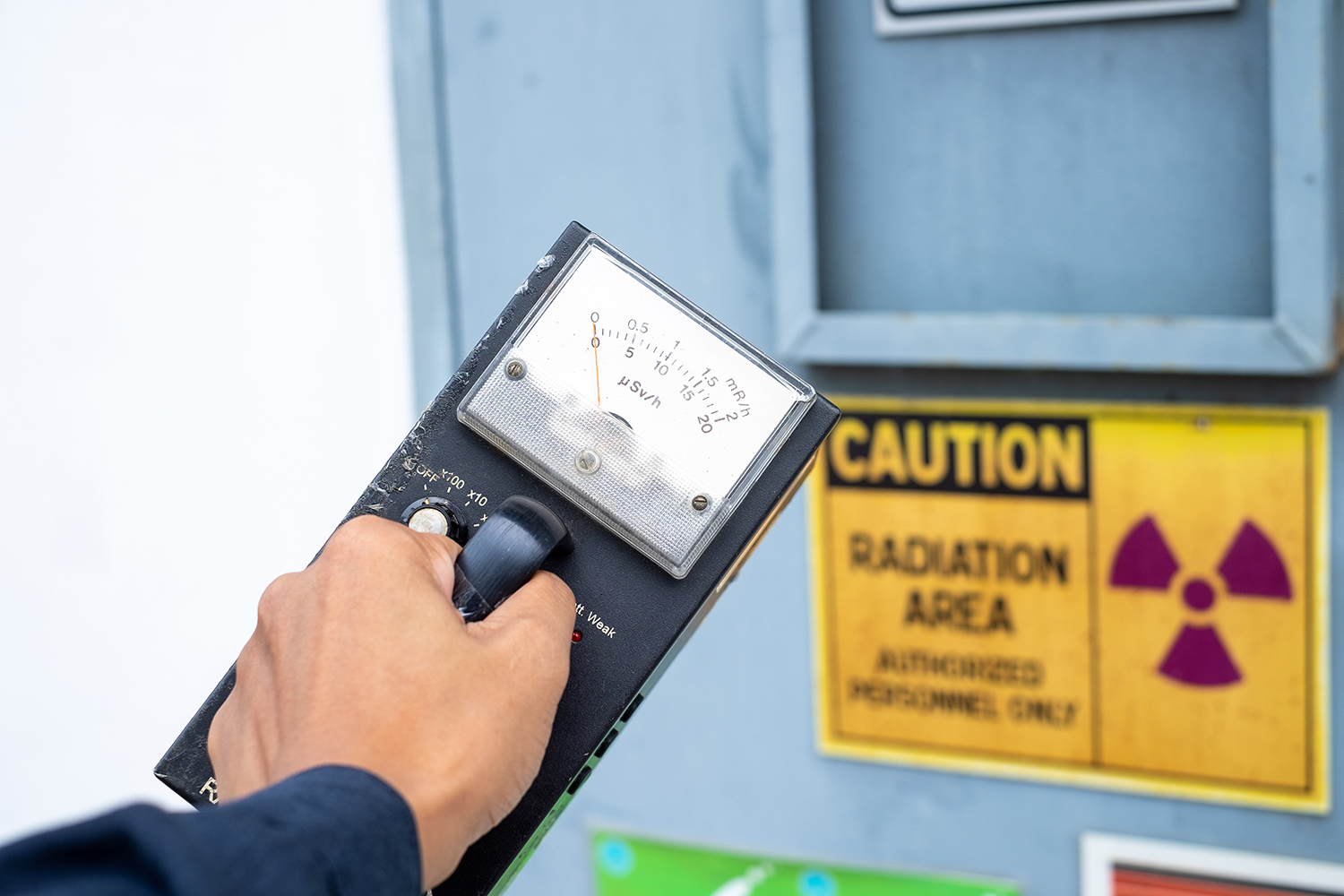
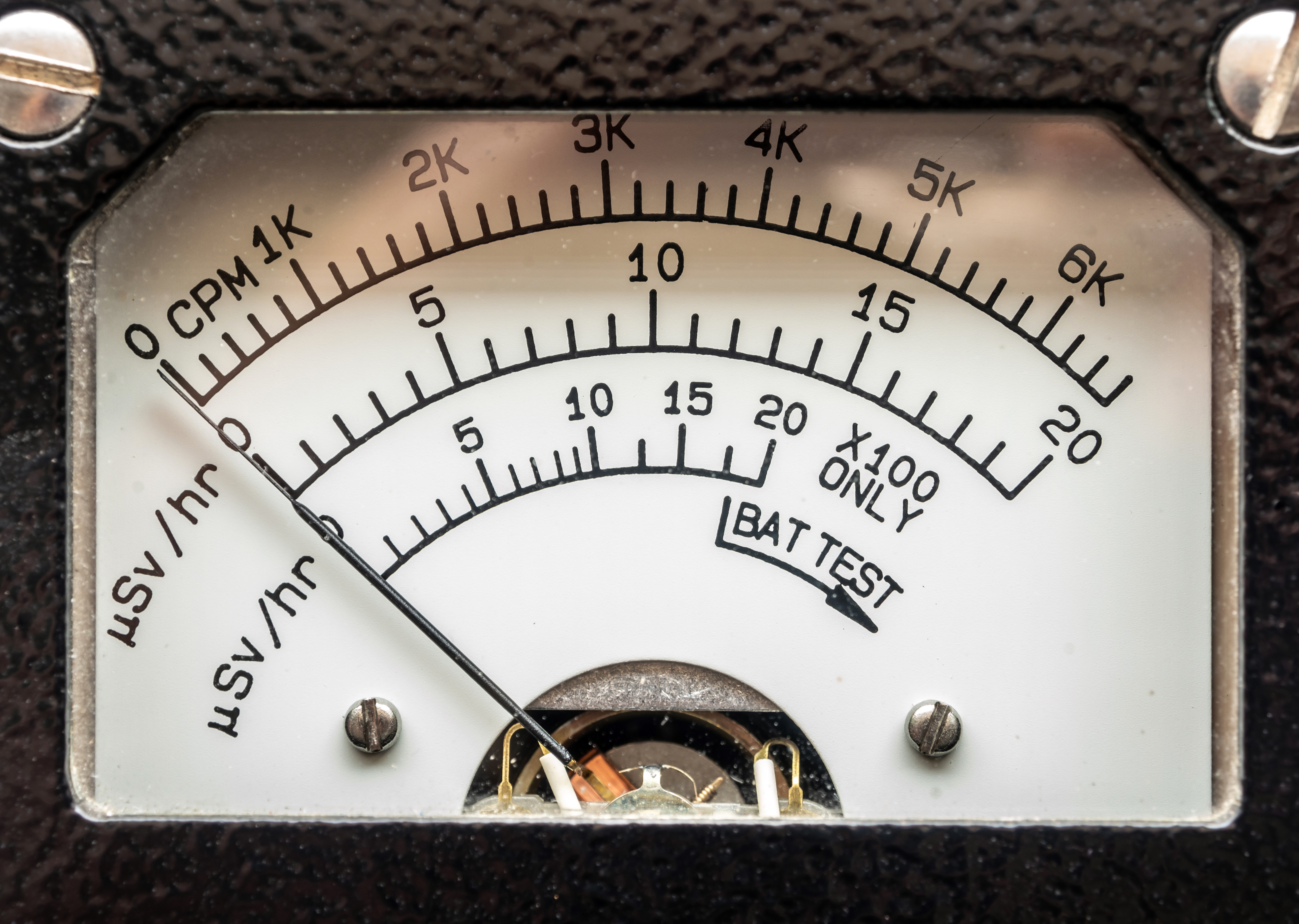
Supervisor use the survey meter to checks the level of radiation

The Units to Measure Radiation: Explained

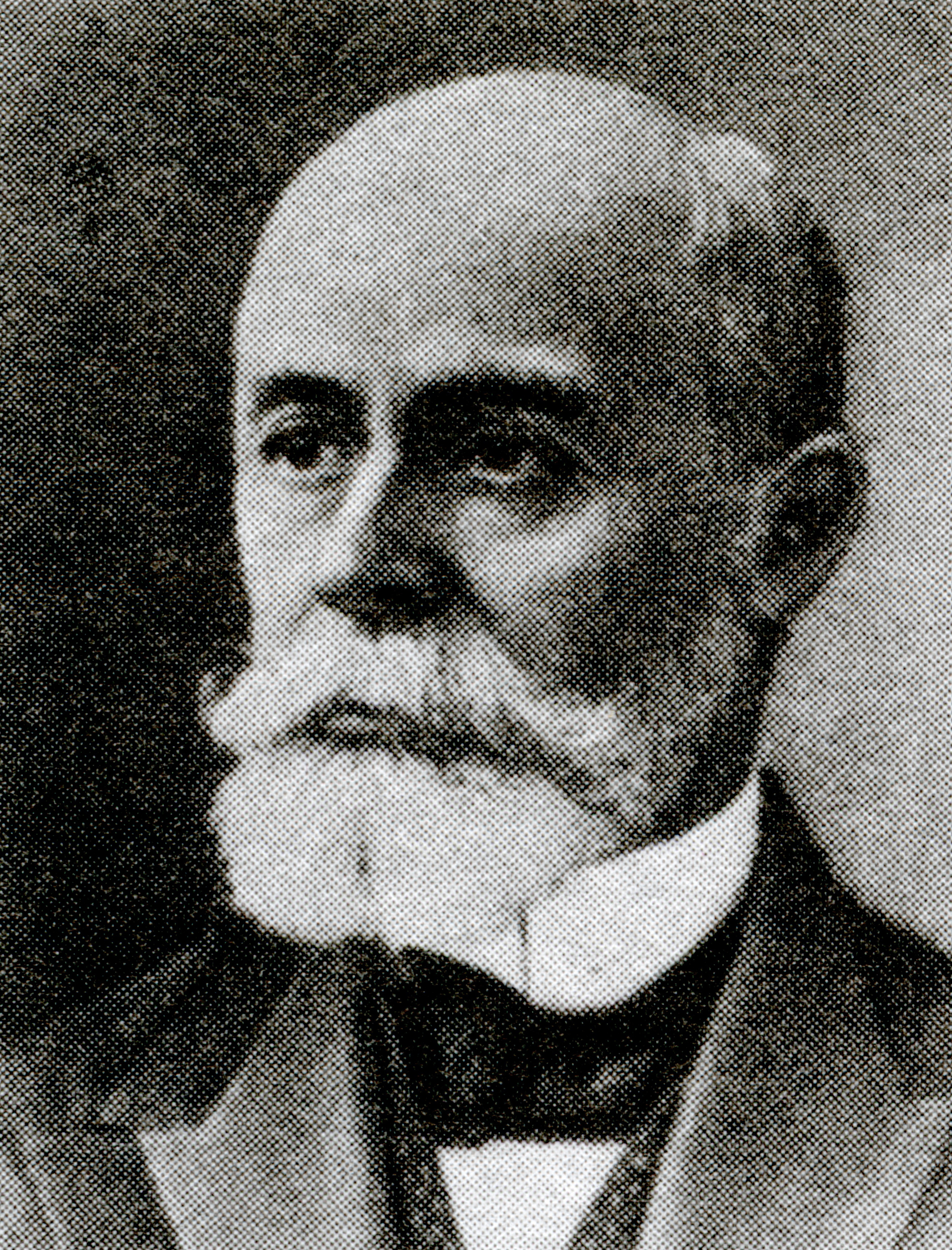
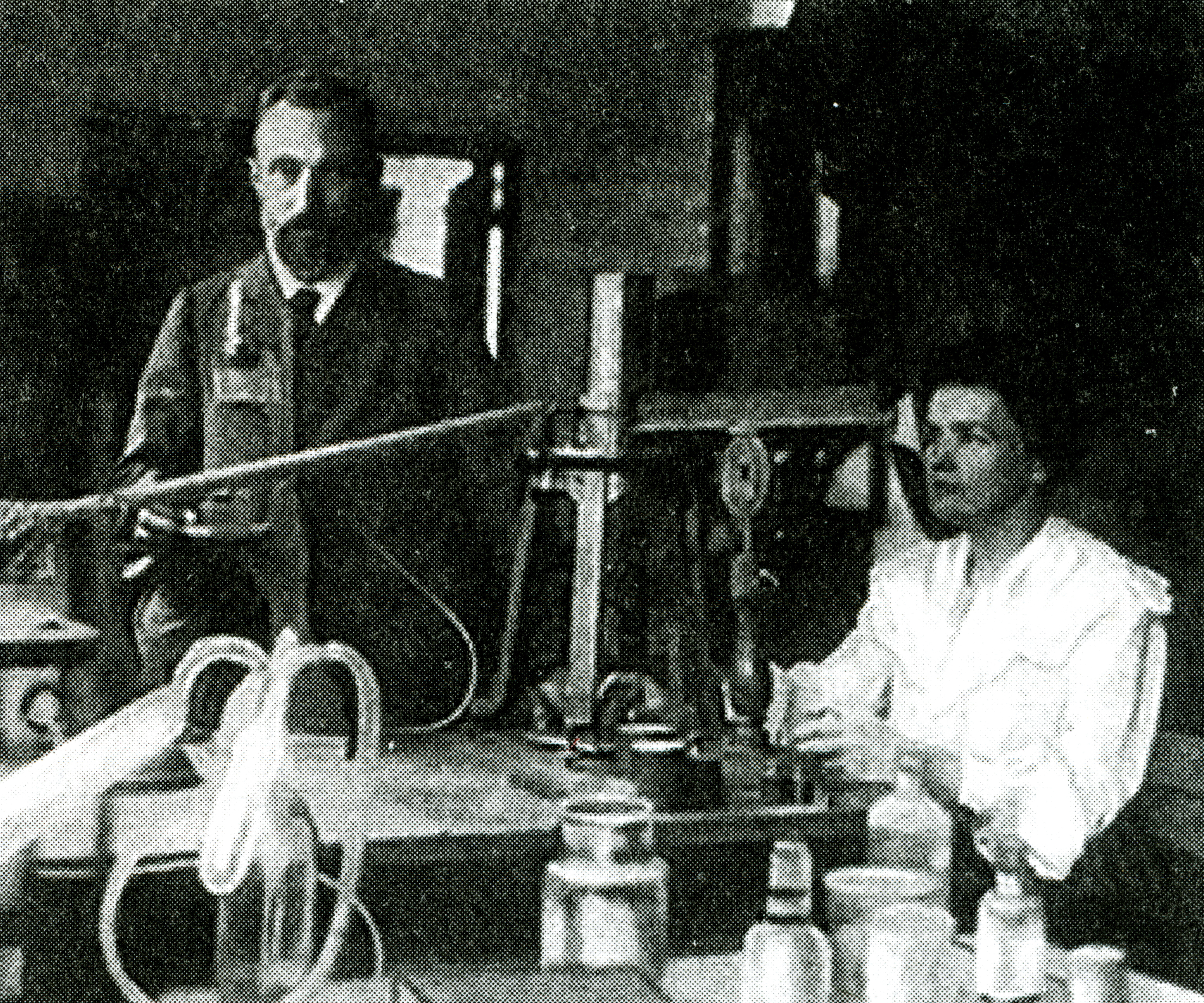
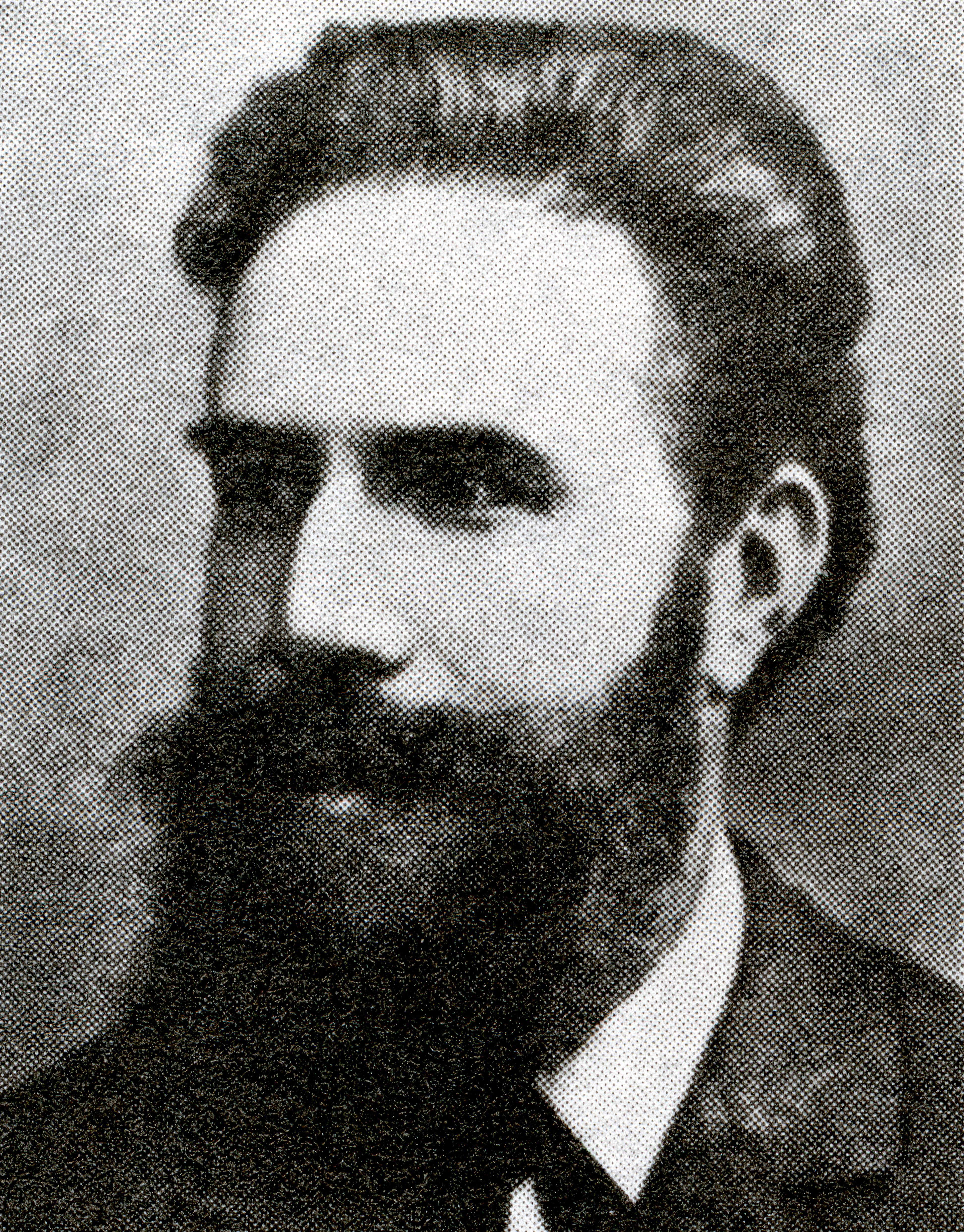
The history of radiation units ties closely to the development of our understanding about radiation and its effects. The discovery of x-rays and radioactivity in the late 19th century by scientists like Wilhelm Roentgen, Henri Becquerel, and Pierre Curie paved the way for the exploration of radiation measurement.

As our knowledge of radiation’s effects on living organisms grew, the need for standardized units became evident. The roentgen was one of the earliest units to measure ionization, followed by the introduction of the curie to measure radioactivity. Over time, advancements in our understanding of radiation’s biological effects led to the development of units like the rem and the sievert.

Creating Radiation Units

The development of the SI system (International System of Units) established a standardized set of units to provide a coherent and universal way to measure radiation. The gray and the sievert were introduced as the primary units for absorbed dose and equivalent dose, respectively, within the SI system.

Four distinct yet interconnected units quantify radioactivity, exposure, absorbed dose, and dose equivalent. The mnemonic R-E-A-D creates a simple way to recall these units, which consist of a combination of commonly used (British, e.g., Ci) and internationally recognized (metric, e.g., Bq) units.1 Below, we will detail the mnemonic and discuss the history of the radiation units along with their relevant scientists:

Radioactivity defines the release of ionizing radiation from a substance. Whether it emits alpha or beta particles, gamma rays, x-rays, or neutrons, the radioactivity of a material is a measure of how many atoms within it decay over a specific period of time. The curie (Ci) and becquerel (Bq) units quantify radioactivity.1


Antoine Henri Becquerel was a French physicist, engineer, and Nobel laureate who discovered evidence of radioactivity. Becquerel’s earliest works centered on the subject of his doctoral thesis: the plane polarization of light, with the phenomenon of phosphorescence and absorption of light by crystals. Early in his career, Becquerel also studied the Earth’s magnetic fields. In 1896, Becquerel discovered evidence of radioactivity while investigating phosphorescent materials such as some uranium salts. For his work in this field, he shared the 1903 Nobel Prize in Physics with Marie Curie and Pierre Curie. The SI unit for radioactivity, becquerel (Bq), is named after him.2

The curie (Ci) unit was created in 1910 by the International Congress of Radiology to measure radioactivity. Pierre Curie, another French physicist, and his wife Marie Curie, who also sat on the committee that named the unit, were the inspirations for the name through their radioactive studies. The original definition of the curie was “the quantity or mass of radium emanation in equilibrium with one gram of radium (element)”.3 In 1975, the becquerel replaced the curie as the official radiation unit in the International System of Units (SI) where 1 Bq = 1 nuclear decay/second4. The relationship between the two units is 1 Ci = 37 GBq (giga becquerels).

Exposure quantifies the extent of radiation going through the atmosphere that reaches a person’s body or a material. Numerous radiation monitors gauge exposure, utilizing the units of roentgen (R) or coulomb/kilogram (C/kg).1

The roentgen is a legacy unit of measurement for the exposure of X-rays and gamma rays. This unit is defined as the electric charge freed by such radiation in a specified volume of air divided by the mass of that air and has the value 2.58 x 10⁻⁴ C/(kg air).5 It was named after Wilhelm Roentgen, a German physicist who discovered X-rays and was awarded the first Nobel Prize in Physics for the discovery.

In 1928, the roentgen became the first international measurement quantity for ionizing radiation defined for radiation protection. This is because it was, at the time, the most easily replicated method of measuring air ionization by using ion chambers.6 However, although this was a major step forward in standardizing radiation measurement, the roentgen had a disadvantage: it was only a measure of air ionization rather than a direct measure of radiation absorption in other materials, such as different forms of human tissue. As a result, it did not take into account the type of radiation or the biological effects of the different types of radiation on biological tissue. Consequently, new radiometric units for radiation protection came to be which took these concerns into account.7

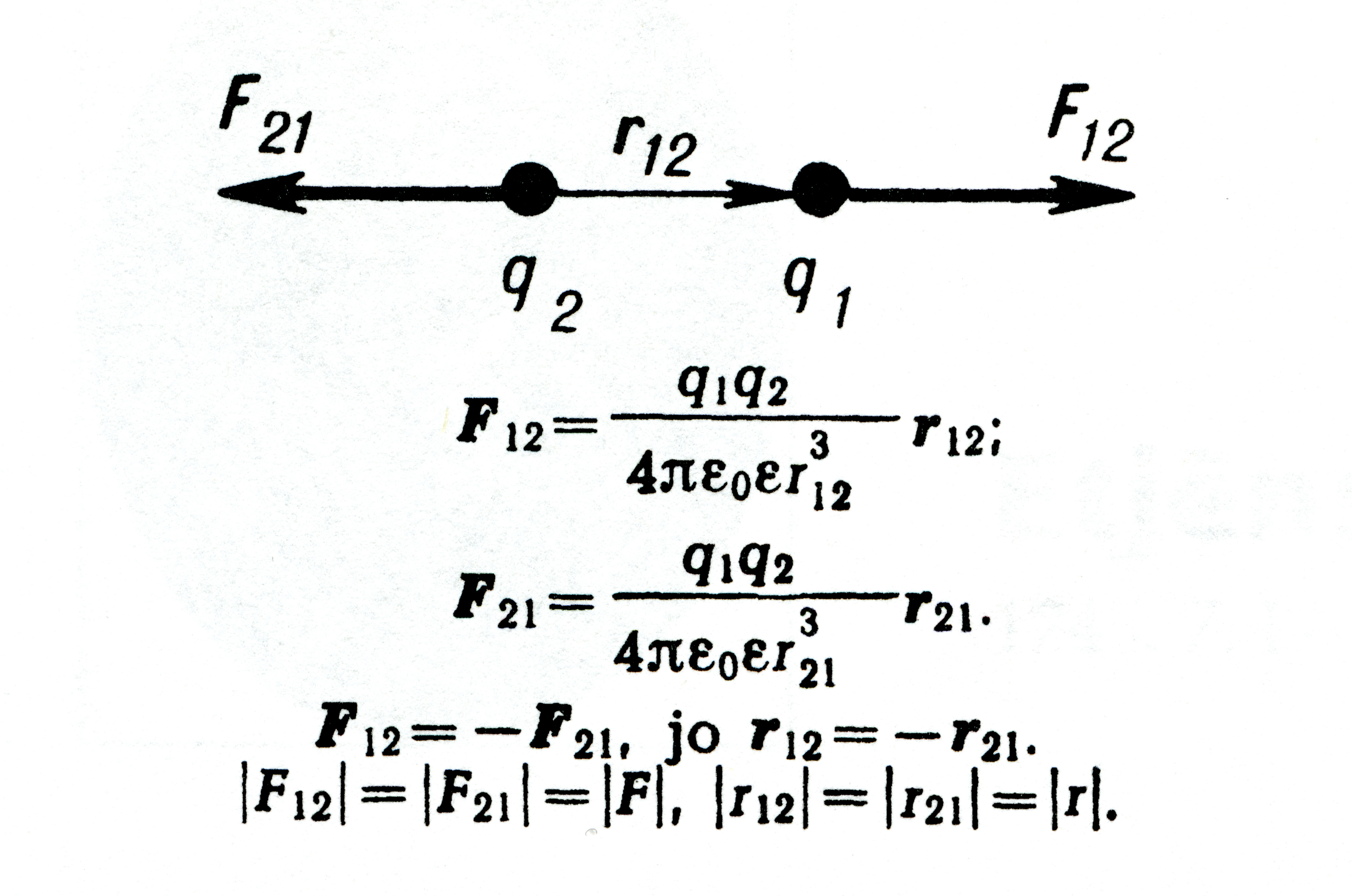
The SI unit for measuring exposure to ionizing radiation is coulomb per kilogram (C/kg). Interestingly, unlike other SI radiation units, this unit does not have a specific name. It officially replaced the previous unit, the roentgen, in 1975, with a transition period of at least ten years.8 The SI unit of electric charge, the coulomb, was named in honor of Charles-Augustin de Coulomb in 1880. Charles-Augustin de Coulomb was a French physicist whose best-known work is his formulation of Coulomb’s law. This law states that the force between two electrical charges is proportional to the product of the charges and inversely proportional to the square of the distance between them. He also made important contributions to the fields of electricity, magnetism, applied mechanics, friction studies, and torsion.9

Absorbed dose refers to the quantity of energy that is absorbed by an object or person where the energy is deposited by ionizing radiation as it passes through materials or the body. The units, radiation absorbed dose (rad) and gray (Gy), measure absorbed dose.1

In 1953, the International Commission on Radiation Units and Measurements (ICRU) adopted the unit rad at the Seventh International Congress of Radiology. This was the unit that replaced the rep, roentgen equivalent physical (detailed later in this blog). Although many believe that the rad is an abbreviation of “radiation absorbed dose”, the ICRU never identified it as such. This suggests that the term “rad” was as a standalone word to be a unit for absorbed dose. There was no documented discussion regarding the use of the rad prior to the Seventh International Congress of Radiology. The closest discussion was during the meeting in 1951 when they determined the need for this type of unit. In 1975, the gray (Gy) replaced the rad as the SI unit of absorbed dose where 1 Gy = 100 rad.10

Louis Harold Gray was a 20th century English physicist who worked mainly on the effects of radiation on biological systems. He was one of the earliest contributors to the field of radiobiology. He worked as a hospital physicist at Mount Vernon Hospital in London and developed the Bragg–Gray equation in collaboration with the father and son team of William Henry Bragg and William Lawrence Bragg. Bragg-Gray theory is the basis for the cavity ionization method of measuring energy absorption by materials exposed to ionizing radiation. Gray’s contributions to radiobiology were numerous. Amongst many other achievements, he developed the concept of RBE (Relative Biological Effectiveness) of doses of neutrons and initiated research into cells in hypoxic tumors and hyperbaric oxygen.11 Gray defined a unit of radiation dosage (absorbed dose) which was later named after him as an SI unit, the gray.

Dose equivalent, also known as effective dose, is a measurement that combines the amount of radiation absorbed and the impact it has on the human body. When it comes to beta and gamma radiation, the dose equivalent is equal to the absorbed dose. However, for alpha and neutron radiation, the dose equivalent surpasses the absorbed dose because these types of radiation have a greater biological impact resulting from their increased ability to damage tissue. To quantify dose equivalent, we use the units of roentgen equivalent man (rem) and sievert (Sv).1

Roentgen equivalent man, or “rem”, was first proposed for use in 1945, but under a different abbreviation. The roentgen was the only unit capable of expressing a radiation exposure at the time. However, it fell short being specifically measurable for photons. Workers came into contact with many other forms of radiation such as alpha particles, beta particles, and neutrons, so Herbert Parker, British-American physicist, created units that would be able to gauge exposures to many types of radiation. These were the roentgen equivalent physical (rep) and the roentgen equivalent biological (reb). Due to similarity in pronunciation between rep and reb, reb was eventually renamed to roentgen equivalent man or mammal (rem) to avoid confusion.

The first appearances of the rem unit in scientific literature were not until 1950.10 The rem related to the rad by multiplying the latter by a quality factor (QF) used to account for the varying biological effects of the different types of radiation. The rad in turn may be obtained from the roentgen by multiplying a dose conversion factor. In air, the dose conversion factor relationship between rad and roentgen is 1 R = 0.88 rad. The absorbed dose to a material is in turn found by multiplying 0.88 by the ratio of the mass energy absorption of the material to that of air.12

Rolf Maximilian Sievert was a Swedish medical physicist. He is best known for his work on the biological effects of ionizing radiation and his pioneering role in the measurement of doses of radiation, especially in its use in the diagnosis and treatment of cancer.13 Sievert contributed significantly to medical physics, earning him the title of “Father of Radiation Protection”. The sievert (Sv), the SI unit representing the stochastic health risk of ionizing radiation, is named after him. The sievert officially replaced the rem as the international SI unit in 1979 with 1 Sv = 100 rem.14

Conclusion

In summary, the history of radiation units is a journey that reflects the progress in our understanding of radiation’s properties and its impact on living organisms. The development of these units has enabled safer and more accurate measurement and assessment of radiation exposure and its effects on human health. By understanding the intricate relationship between radiation and our bodies, we can now take proactive measures to mitigate its harmful effects and promote a safer environment for all.

Sources

  1. NRC: Measuring Radiation. Nrc.gov. Published 2017. https://www.nrc.gov/about-nrc/radiation/health-effects/measuring-radiation.html
  2. The Nobel Prize. The Nobel Prize in Physics 1903. NobelPrize.org. Published 2019. https://www.nobelprize.org/prizes/physics/1903/becquerel/biographical/
  3. Curie – Unit of Radioactivity | nuclear-power.com. Nuclear Power. https://www.nuclear-power.com/nuclear-engineering/radiation-protection/units-of-radioactivity/curie-unit-of-radioactivity/
  4. ‌ Bell DJ. Becquerel (SI unit) | Radiology Reference Article | Radiopaedia.org. Radiopaedia. https://radiopaedia.org/articles/becquerel-si-unit
  5. ‌ Bashir U. Roentgen (unit) | Radiology Reference Article | Radiopaedia.org. Radiopaedia. Accessed August 29, 2023. https://radiopaedia.org/articles/roentgen-unit?lang=us
  6. ‌ Roentgen – Unit of Exposure | nuclear-power.com. Nuclear Power. Accessed August 29, 2023. https://www.nuclear-power.com/nuclear-engineering/radiation-protection/radiation-exposure/roentgen-unit-of-exposure/
  7. ‌ Roentgen (unit) explained. everything.explained.today. Accessed August 29, 2023. http://everything.explained.today/Roentgen_(unit)/
  8. ‌ Bell DJ. Coulomb per kilogram | Radiology Reference Article | Radiopaedia.org. Radiopaedia. Accessed August 29, 2023. https://radiopaedia.org/articles/coulomb-per-kilogram
  9. ‌ Laboratory NHMF. Charles-Augustin de Coulomb – Magnet Academy. nationalmaglab.org. https://nationalmaglab.org/magnet-academy/history-of-electricity-magnetism/pioneers/charles-augustin-de-coulomb/
  10. ‌ Why Did They Call It That? The Origin of Selected Radiological and Nuclear Terms. Museum of Radiation and Radioactivity. Accessed August 29, 2023. https://orau.org/health-physics-museum/articles/selected-radiological-nuclear-terms.html#rad
  11. ‌ LH Gray Memorial Trust: About L.H. Gray. www.lhgraytrust.org. Accessed August 29, 2023. http://www.lhgraytrust.org/lhgraybiography.html
  12. ‌Dosimetric Quantities and Units. U.S. NRC. Published October 25, 2010. Accessed September 11, 2023. https://www.nrc.gov/docs/ML1122/ML11229A688.pdf
  13. Aip.org. Published 2023. Accessed August 29, 2023. https://pubs.aip.org/physicstoday/Online/8433/Rolf-Sievert
  14. ‌ Bell DJ. Sievert (SI unit) | Radiology Reference Article | Radiopaedia.org. Radiopaedia. https://radiopaedia.org/articles/sievert-si-unit?lang=us
21 Apr 2021

Deterministic vs. Stochastic Effects: What Are the Differences?

Ionizing radiation is useful for diagnosing and treating a range of health conditions–broken bones, heart problems, and cancer, for example.  Medical imaging with x-rays, diagnostic radiopharmaceuticals, and radiation therapy are often life-saving procedures.

However, the accidental or misuse of medical radiation can sometimes cause unforeseen and unfortunate consequences.  Radiation protection guidelines and policies help to ensure the safe use of radiation in the medical setting for both patients and staff.

The health effects of ionizing radiation are usually classified into two categories: deterministic and stochastic.

Deterministic Effects


According to the International Atomic Energy Agency (IAEA), a health effect that requires a specific level of exposure to ionizing radiation before it can occur is called a deterministic effect. The severity of a deterministic effect increases as the dose of exposure increases and considers a minimum threshold, below which no detectable clinical effects occur. This type of effect is predictable and reproducible.  For example, localized doses to certain parts of the body at increasing levels will result in the same biological effects.

Deterministic effects are caused by severe cell damage or death. Individuals who experience the physical effects of this cell death do so when it is large enough to cause significant tissue or organ impairment.

Deterministic effects are short-term, adverse tissue reactions resulting from a dose that is significantly high enough to damage living tissues.  The severity of a deterministic effect increases with radiation dose above a threshold, below which the detectable tissue reactions are not observed. 

Deterministic effects are usually predictable and reproducible.  For example, localized doses to certain parts of the body at increasing levels will result in well-understood biological effects.

how to understand and communicate radiation risk diagram
Figure 1 Radiation – Deterministic and Stochastic Effects – Image Wisely, March 2017 “How to Understand and Communicate Radiation Risk”

Some examples of deterministic effects include:

  • Radiation-induced skin burns
  • Acute radiation syndrome
  • Radiation sickness
  • Cataracts
  • Sterility
  • Tumor Necrosis

Stochastic Effects


Stochastic effects are probabilistic effects that occur by chance.  An extremely rare stochastic effect is the development of cancer in an irradiated organ or tissue.  The probability of occurrence is typically proportional to the dose received. Stochastic effects after exposure to radiation occur many years later (the latent period).  The severity is independent of the dose originally received.

Since many agents in the environment are also known carcinogens, and since many cancers occur spontaneously, it is not possible in most cases to directly link radiation exposure to an observed cancer.  If a population group receives a dose of ionizing radiation at one time, it is therefore not possible to predict who in that group will develop cancer, if any, or to tell if the people who do develop cancer did so as a result of the dose of ionizing radiation or some other lifestyle factor, such as smoking.   

Examples of stochastic effects include:

  • Cancer
  • Heritable or genetic changes


Dose Limits and Radiation Protection


In our day-to-day lives, we are exposed to both background and man-made sources of radiation.  Everyone receives radiation exposure from natural cosmic and solar rays, and radionuclides in soil.  The benefits of diagnostic and therapeutic medical radiation far exceed the risks.  Fortunately, the health risks associated with natural background levels are small, and by regulations, we are protected from man-made radiation. 

The National Council on Radiation Protection and Measurements (NCRP) recommends dose limits for managing exposures to ionizing radiation and protecting humans from adverse effects.  Their purpose is to prevent acute and chronic radiation-induced tissue reactions (deterministic effects) and to reduce the probability of cancer (stochastic effect) while maintaining the benefits to people and society from activities that generate radiation exposures (NCRP Report No. 180, 2018).

Type of limit Radiation worker Public
Stochastic limits Effective dose, whole body (mSv/year) 50 1
Deterministic limits Tissue absorbed dose (mGy/year)
Lens of the eye 50 15
Skin 500
Extremities (hands and feet) 500

Figure 2.  Values from NCRP Report No. 180, Management of Exposure to Ionizing Radiation:  Radiation Protection Guidance for the United States (2018).

The concept of dose limits also takes into account the ideas that any use of radiation should do more good than harm, and that permissible exposure should be maintained “as low as reasonably achievable” (ALARA).   In line with this philosophy, medical professionals strive to minimize medical radiation exposures to patients without compromising imaging quality and therapy effectiveness. 

Conclusion


Adverse health effects can occur after exposure to ionizing radiation.  For radiation protection, scientific advisory organizations have recommended dose limits to prevent deterministic effects and reduce the probability of stochastic effects in radiation workers, medical professionals, patients, and other members of the general public. 


Versant Physics is a full-service medical physics and radiation safety consulting company based in Kalamazoo, MI. Contact us for all of your regulatory, radiation safety, and personnel dosimetry needs.

Sources:

  1. https://hps.org/publicinformation/ate/faqs/regdoselimits.html
  2. https://www.nrc.gov/reading-rm/basic-ref/glossary/non-stochastic-effect.html
  3. https://www.nrc.gov/about-nrc/radiation/around-us/uses-radiation.html
  4. https://www.radioactivity.eu.com/site/pages/Deterministic_Effects.htm
  5. https://www.imagewisely.org/Imaging-Modalities/Computed-Tomography/How-to-Understand-and-Communicate-Radiation-Risk
  6. https://www.radiation-dosimetry.org/what-is-dose-limit-radiation-definition/