
The idea that radiation exposure is inescapable through everyday life might cause a feeling of concern or even trepidation. To make it more palatable to the public, a somewhat quirky unit of measurement was born for absorbed radiation dose. This is the “Banana Equivalent Dose”, or BED (not to be confused with the other well-known BED, the Biologically Effective Dose). Bananas, a fruit found in many homes, all contain a very small but measurable dose of ionizing radiation. The BED provides an opportunity to bring an unusual yet helpful perspective towards discussions on radiation exposure so that the science behind radiation feels more accessible and less mysterious. Let’s explore the BED, why radiation in food exists, and what it teaches us about daily radiation encounters.
What is the Banana Equivalent Dose?
The Banana Equivalent Dose (BED) is an informal unit of measurement that expresses a radiation dose in terms of the amount one person would receive from eating a single banana.1 Instead of the knowledge that radiation can exist in our food being a cause for concern, it provides a chance for better understanding of a difficult topic. The idea of the BED offers a way for the public to frame seemingly large or unfamiliar numbers relating to ionizing radiation into a more meaningful context. Although it isn’t an official unit endorsed by regulatory agencies or scientific organizations, the BED has shown to be an easier baseline for regular citizens to comprehend than attempting to explain rems or sieverts.
The average BED is about 0.1 microsieverts (μSv) per banana (1 μSv = 0.000001 sieverts, a recognized unit for measuring ionizing radiation dose).1 For some context, an average person will receive roughly 2,000 to 3,000 μSv per year due to natural background radiation like cosmic rays, radon gas, and terrestrial sources. This is equivalent to 20,000 to 30,000 bananas. When someone consumes a single banana, a minuscule addition to this background radiation occurs.
Why Are Bananas Radioactive?
Bananas are well-known for being rich in potassium, which is an essential mineral for human nutrition. They gain most of this potassium through the uptake of the mineral via the roots of their trees. There is also deposition, which is when the fruit absorbs radioactive particles in the air that settle on it.2 Naturally occurring potassium will normally be comprised of the mostly stable isotope potassium-30, but close to 0.012% of the potassium is the radioactive isotope, potassium-40. Potassium-40 decays with a half-life that equals about 1.3 billion years which means it releases radiation at a very slow rate.3
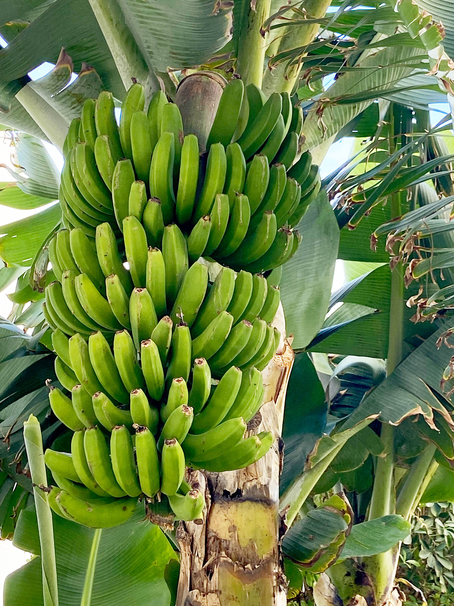
After eating a banana, both radioactive and non-radioactive potassium isotopes enter your body, becoming part of the natural chemical makeup. The human body strictly regulates potassium levels, so any excess is excreted fairly quickly. After some time to digest and excrete, your body is able to return your internal dose of potassium-40—including its radiation—to its equilibrium, regardless of whether you might eat one banana or even a dozen.3
How the Banana Equivalent Dose Came to Be
The true origin of the Banana Equivalent Dose is not clearly recorded in any sources. However, it is reasonable to surmise that the unit was suggested during research on radiation found in foods. The earliest known mention of the BED is from 1995 on a recorded email chain from Gary Mansfield. An employee at Lawrence Livermore National Laboratory at the time, Gary suggests that he found the BED to be “very useful in attempting to explain infinitesimal doses (and corresponding infinitesimal risks) to members of the public”.4
By comparing common procedures like a typical chest X-Ray (about 1000 BEDs) or actions like flying from New York to London (about 400 BEDs) to the dose from a banana, experts can create an easier connection of these common, low-level exposures to the overall idea of radiation safety. Even large organizations like the United States Environmental Protection Agency (EPA) reference the BED in public educational articles.2
The BED has remained a popular option for comparison in scientific forums and educational websites due to familiarity and relatability. Radiation is given a more understandable setting within bananas. The common household snack serves as a reminder that sometimes exposure is not as disastrous as previously believed.
Understanding Radiation Dose with the BED
An issue with radiation being an enigma for many is the misconception that all radiation is harmful, regardless of dose. There are many circumstances where that is not the case. The BED assists with putting radiation exposure into a new light:
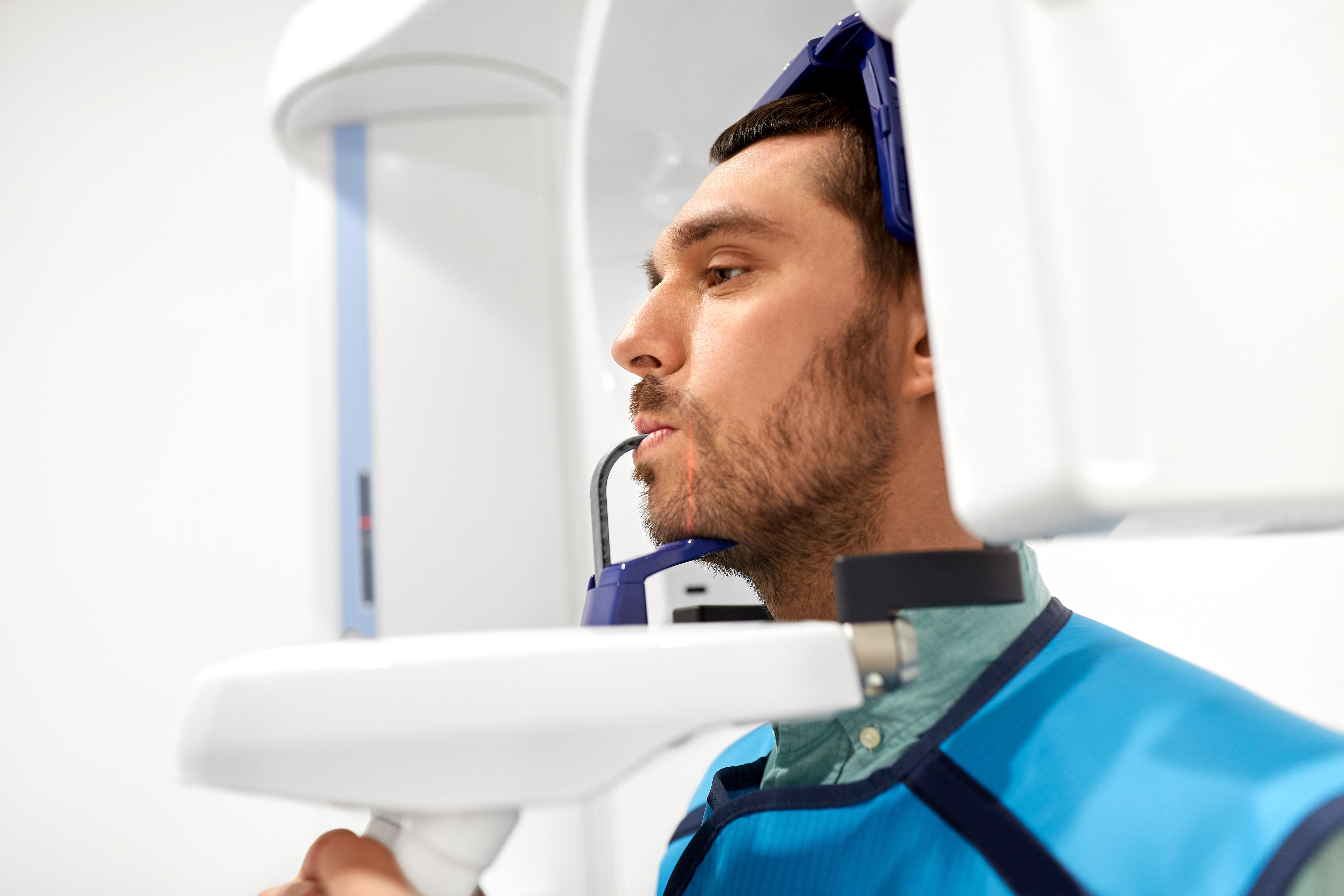
- Dental X-ray: Roughly equivalent to 50 BEDs
- Yearly dose from living near a nuclear power plant: 1 – 100 BEDs
- Chest CT scan: 70,000 BEDs
- Typical targeted dose used in radiotherapy (one session): 20,000,000 BEDs1
Although it may be hard to fully imagine a stack of twenty million bananas, it proves to be a more tangible idea to promote informed understanding of the amount of radiation exposure in a procedure than the more abstract measurements of sievert values for the public.
What Other Foods with Radiation Exist?
Many foods naturally absorb radioisotopes from the soil and water in which they grow, putting bananas into some popular company for regular consumption. Here are some of the most recognizable foods with radioactive elements:
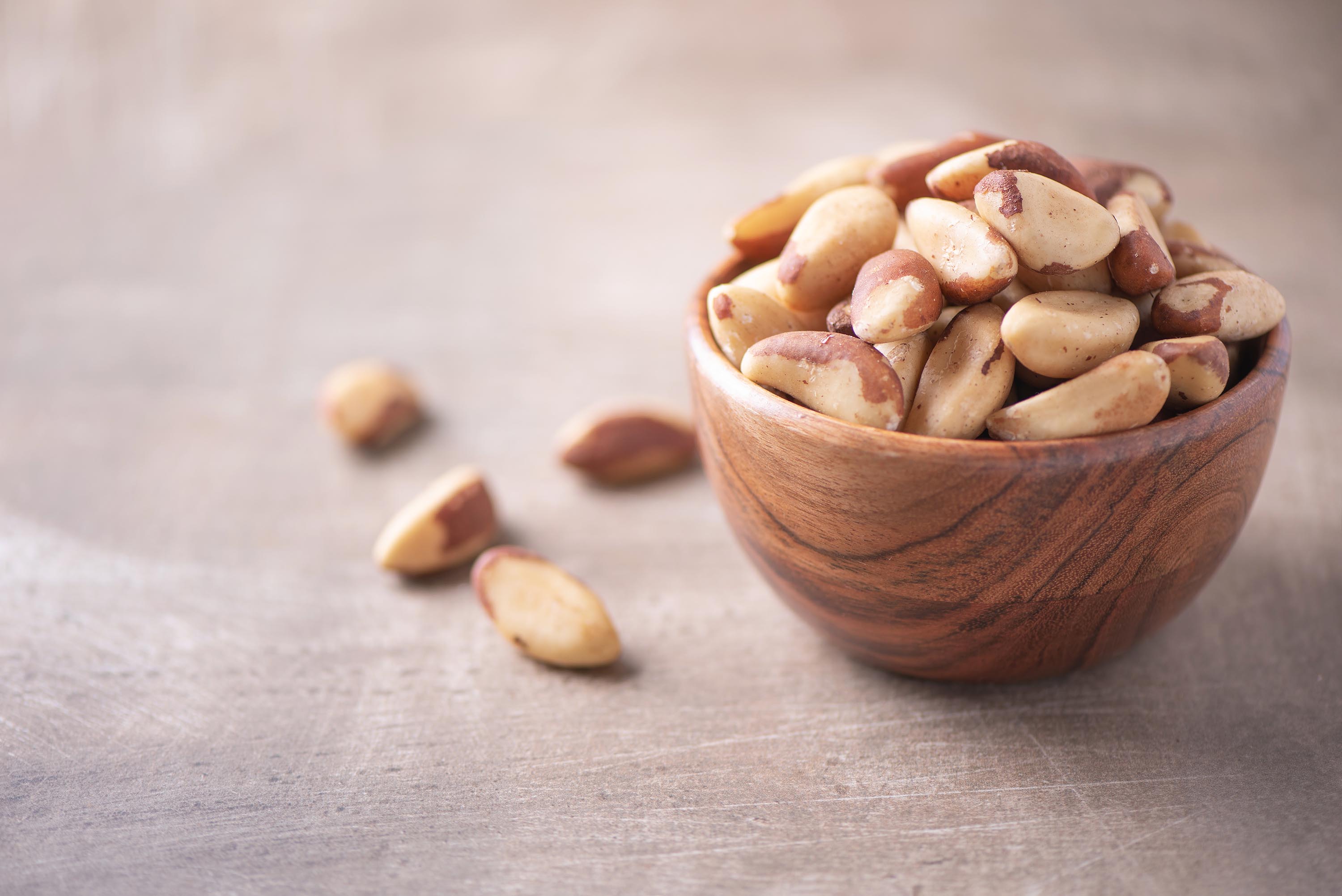
- Brazil nuts are well-known for having high levels of radium-226 and radium-228, with doses that can, at times, be even higher than bananas. This is because Brazil nut trees have deep roots that access radium-rich soil and lead to these concentrations.
- Potatoes are another potassium-rich food and contain potassium-40. Unlike bananas, however, they also absorb small amounts of uranium and thorium.
- Carrots and red meat can accumulate some minor amounts of radioisotopes, particularly potassium-40. However, their doses are minimal compared to annual background radiation.
- Some beer has trace amounts of potassium-40—not unlike other plant-derived foods.5
Of course, it is important to note that, despite the radioactivity in these foods, they pose no health risks when considering consumption in a normal diet. As previously mentioned, the human body is capable of handling low doses of naturally occurring radiation. Regulatory agencies also take care to monitor food safety to safeguard public health.
Conclusion: The Public Value of the BED
When it comes to encouraging better understanding of a complex subject like radiation exposure, there’s value in linking it to a familiar concept in a person’s life. Not only does the BED provide a light-hearted opportunity to understand your day-to-day exposure but it also helps show that consuming food with absorbed radioactive elements does not normally pose a risk to one’s health or make a person radioactive—far from it, even. The Banana Dose Equivalent gives the public a dose measurement that “peels” back the layers of uncertainty to see that radiation is a natural part of the world that’s findable in the food we eat and even ourselves.
Sources
- P2 Nuclear and Par-Cle Physics –Dan Protopopescu. https://www.ppe.gla.ac.uk/~protopop/teaching/NPP/P2-NPP.pdf
- Natural Radioactivity in Food. US EPA. Published November 27, 2018. https://www.epa.gov/radtown/natural-radioactivity-food
- Schwarcz J. McGill University. Office for Science and Society. Published December 30, 2018. https://www.mcgill.ca/oss/article/you-asked/it-true-banana-radioactive
- Banana Equivalent Dose. Iit.edu. Published 2025. http://health.phys.iit.edu/extended_archive/9503/msg00074.html
- Helmenstine A. These 10 Common Foods Are Radioactive. ThoughtCo. Published 2014. https://www.thoughtco.com/common-naturally-radioactive-foods-607456