Category: Radiation Safety

04 Nov 2025
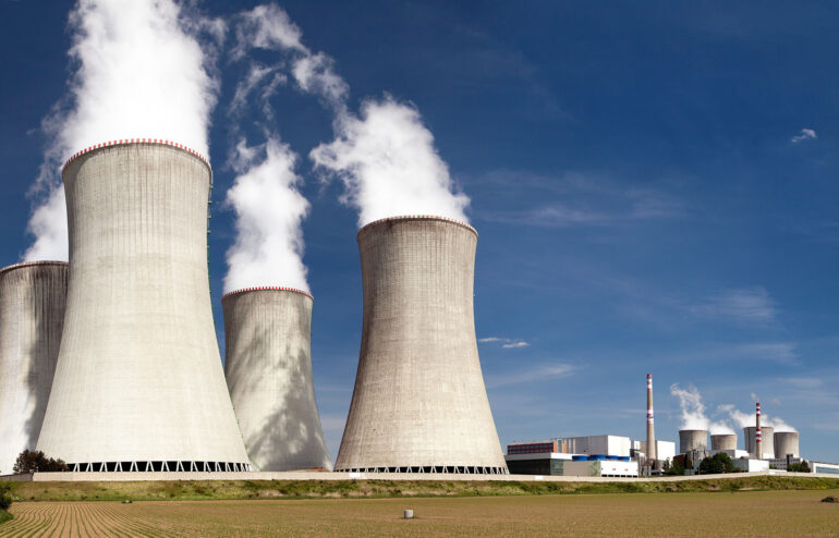
A depiction of a nuclear power plant and its cooling towers.

 The Resurgence of Nuclear Energy

The world is in a new age of change and challenge with developments in technology, climate change, and available resources. Demands for further consideration on renewable energy options are rising, and with it comes a returning interest in nuclear energy. We can see the resurgence of nuclear energy in Versant Physics’ own home state: the Palisades Nuclear Power Plant, which stopped commercial operation in 2022, is being restored through a collaborative effort of Siemens Energy and Holtec International.1

These two companies aim to enhance safety, reliability, and efficiency through their efforts to modernize and renew the Palisades to generate more than 800 megawatts (MW) of electricity.2 The goal for this energy is to power around 800,000 households. Other major companies are pursuing similar restoration projects across the United States for different reasons. Let’s explore the history of nuclear energy, its fall out of favor, and why we’re seeing attention returning to the power source.

The Rise in Nuclear Reactor Development

The first technologies for nuclear reactors and creating nuclear energy came about in the 1930s and 1940s. Its potential as a clean energy source, however, was overshadowed by government demand for nuclear bomb research for World War II. It was not until the war came to an end that a renewed focus on nuclear energy could occur. The sheer capacity of nuclear power had been demonstrated in some of the most devastating ways, but a determination to harness it for good by way of creating steam and electricity rose in the 1950s.3

The first nuclear reactor to ever produce electricity was a small reactor created in the United States by Argonne National Laboratory in 1951. Seeing the rise in potential for power generation, President Eisenhower proposed an “Atoms for Peace” program in 1953. This reoriented research effort contributed significantly towards electricity generation and inspired civil nuclear energy development in the USA.  Great progress within the United States and the rest of the world continued throughout the 1950s with the creation of new reactor designs. These included fast breeder reactors (FBRs), which produce more fissile materials than they consume and are designed to extend nuclear fuel supply to generate electricity, as well as pressurized water reactors (PWRs), a reactor originally designed for the U.S. Navy that uses pressurized water as a coolant and neutron moderator.4,5

Commercial nuclear energy reactors started appearing in 1959 in France, quickly followed by the U.S., the United Kingdom, and Russia across the 1960s.  Many of these reactors were light water designs (either PWRs or boiling water reactors, BWRs) and some could generate up to 250 MW of energy. Within the early 1970s, the world saw its first high-power channel reactors that could generate 1,000 MW of energy.3 Despite these rapid developments of increasing energy outputs, the demand for this new source of power was short lived.

Why the Quick Decline to Nuclear Energy Interest?

Just as nuclear energy orders started coming in during the 1960s, the United States and other countries creating nuclear power plants began to see a decline just as quickly in the 1970s and through the rest of the 20th century.  This was due to unresolved concerns about the latest world war and unforeseen incidents in the future.

The Anti-Nuclear Movement and the Energy Crisis of the 1970s

 Within the midst of the Cold War where the world’s superpowers were locked in their nuclear arms race, animosity towards nuclear products spread throughout the public perspective. Local and national protests grew against the use of any nuclear weaponry or power plants due to the proven potential for destruction from World War II and the looming threat created by the Cold War.6 Producers of these plants were forced to bring production to a slowdown as previous orders from the 1960s were cancelled and interest in any new requests waned.3

In 1973 during the Yom Kippur War, the Arabian state members of the Organization of Petroleum Exporting Countries (OPEC) initiated an embargo on oil exports to the U.S. This created the oil crisis from 1973 which caused the U.S. government to scramble for alternative energy sources. This led to nuclear energy coming back on the table as an option for power, but concerns surrounding the consequences of power plants had only worsened. Within the U.S., many Americans were against storage of radioactive material and waste generated from the nuclear power plants. Protests cited potential danger to the health of residents living near the sites and the negative environmental impact if any waste leaked out of containment.6

The Chernobyl, Three Miles Island, and The Fukushima Disaster Incidents

After the rise of the Anti-Nuclear Movement, incidents relating to established nuclear power plants began to occur. The first recorded nuclear power plant incident happened in 1979 at the Three Mile Island nuclear power plant in the USA. Due to a cooling malfunction, part of the core in Unit 2 of the plant melted, destroying the reactor. Although there were no injuries or adverse health effects from the event, the event caused widespread confusion and concern about the impact of the incident.7

The Chernobyl disaster occurred in 1986 and is notably the worst incident to have occurred in history. This nuclear power plant is in Chernobyl, Ukraine, and a sudden surge of power during a reactor systems test destroyed Unit 4 of the station. Investigation of the incident determined that a lack of proper safety culture caused the devastation at the Chernobyl power plant8. Many site workers were killed or had acute radiation sickness.9

Image depicting a full view of the Chernobyl nuclear power plant.

These incidents only heightened anti-nuclear movements in the end of the 20th century. Due to the strong reluctance towards increasing nuclear energy production, new facilities were rare into the early 2000s.  The Fukushima Daiichi accident of 2011, in which an unprecedented natural disaster hit Japan and overpowered the outdated tsunami countermeasures of the Daiichi site, continued to incite trepidation towards the safety of nuclear power plants.10 The world is seeing more alternative energy sources in response to climate change, but through solar, wind, and geothermal routes. Nuclear energy produces only 9% of the world’s electricity as of 2025,11 but things may change soon. After over a decade without serious troubles, eyes are returning to nuclear energy as power for new technologies.

Nuclear Energy’s New Appeal

One of the biggest technological advancements that the world is adapting to is the rise in artificial intelligence (AI). Companies across the globe are implementing their own AI systems that require very large, energy demanding data centers to function. For one request to ChatGPT, the International Energy Agency (IEA) has determined that the response the AI provides requires ten times more electricity than someone completing a Google search.12 Current power grids are struggling to keep up with the demands of artificial intelligence. In response, companies creating AI who need to maintain these data centers are casting attention towards untapped nuclear energy sources.

Who are some of the big names looking to harness nuclear energy?

One of the first deals made that suggested a reemergence of nuclear energy was in September of 2024. The tech giant Microsoft signed an agreement with Constellation Energy to restart the Unit 1 nuclear reactor at the Three Mile Island plant in Pennsylvania.13 Once the site is up and running, Microsoft hopes to have 835 megawatts (MW) of new power they can dedicate to their data centers. Whether this $1.6 billion deal is worth its steep price tag depends on time—the Three Mile Island plant originally closed in 2019 due to economic challenges. Now, Microsoft faces getting the nuclear reactors back into working order, renewing its operating licenses, and starting operations again by 2028.13

The social media and technology giant, Meta, aims to take a similar approach to Microsoft for their AI power needs. By partnering with Constellation Energy, Meta hopes to access 1,121 MW of nuclear energy through an established Illinois nuclear facility. This will bolster the electricity needs for Meta to continue its AI developments. They hope to increase data centers and important hubs for tasks like “AI model training, content delivery, and cloud services”.14

While Microsoft and Meta will be taking advantage of established nuclear power plants, Google and Amazon are approaching their move into nuclear energy differently. These companies are looking to invest in the creation of small modular reactors (SMRs). This type of plant currently doesn’t operate anywhere in the United States and only a few exist in the world. SMRs produce less than 300 MW per reactor. Amazon has made a deal with X-Energy, a company capable of designing “high-temperature gas reactors,” and Energy Northwest. They aim to create four reactor modules that would provide at least 320 MW combined.14 Meanwhile, Google has moved into an agreement with Kairos, a company that develops “molten salt-cooled, TRISO fuel-powered” reactors. The goal of power production from these future SMRs for Google would be 500 MW by 2035.14

Will nuclear energy be the long-term solution for AI needs?

The unexpected energy demand for artificial intelligence is driving these trillion-dollar companies to invest in nuclear energy potential, but will this be a long-term investment? Some suggest that the eventual solution to this power-hungry technology will be the technology itself. Artificial intelligence is continuously growing, being trained to learn more and do more. Some believe that AI algorithms will eventually become the best path to determining its own energy management. This would be achievable through its capacity to “identify patterns in data, detect anomalies, and anticipate and forecast future results”.12

Given enough time to “self-reflect”, AI could determine how to optimize its own functioning to reduce its carbon footprint and power necessary to be sustained. In the meantime, nuclear power plants will be giving these tech giants an opportunity to obtain the energy they need. With the increasing pressure of honoring climate commitments and lowering greenhouse gases, they may also find that nuclear energy suits more than just their AI needs.

Conclusion

Nuclear energy is still an industry that is seen first for the disastrous events from the past and their repercussions. Power plants are considered as hazardous potential for nuclear waste leaks, core meltdowns, and a threat for radiation exposure to those who live nearby. However, as the need for cleaner energy increases in demand, we may find that nuclear is an option to reconsider.

Handling of radiation and nuclear power has improved over recent decades. Now more than ever, safety concerns are taken seriously to avoid repetitions of historical incidents. With investments from large companies like Microsoft and Meta, the resurgence of nuclear energy could provide a path to expanded opportunities of clean power provision for more than just AI data centers. By focusing on safety, responsible handling, and sustainability as core features of future nuclear power plants, perhaps the true potential for nuclear energy will be revealed.

Sources

  1. Palisades Nuclear Power Plant. Siemens-energy.com. Published 2025. Accessed October 30, 2025. https://www.siemens-energy.com/global/en/home/references/recommissioning-palisades-nuclear.html
  2. Davidson K. Palisades Nuclear Plant on Lake Michigan shoreline one step closer to reopening • Michigan Advance. Michigan Advance. Published July 25, 2025. Accessed October 30, 2025. https://michiganadvance.com/briefs/palisades-nuclear-plant-on-lake-michigan-shoreline-one-step-closer-to-reopening/
  3. ‌World Nuclear Association. Outline History of Nuclear Energy. world-nuclear.org. Published May 2, 2024. https://world-nuclear.org/information-library/current-and-future-generation/outline-history-of-nuclear-energy
  4. Breeder reactor – Energy Education. Energyeducation.ca. Published 2017. https://energyeducation.ca/encyclopedia/Breeder_reactor
  5. World Nuclear Association. Nuclear Power Reactors. world-nuclear.org. Published January 23, 2025. https://world-nuclear.org/information-library/nuclear-fuel-cycle/nuclear-power-reactors/nuclear-power-reactors
  6. Antinuclear movement of the 1970s | EBSCO. EBSCO Information Services, Inc. | www.ebsco.com. Published 2022. https://www.ebsco.com/research-starters/history/antinuclear-movement-1970s
  7. World Nuclear Association. Three Mile Island Accident – World Nuclear Association. world nuclear association. Published October 11, 2022. https://world-nuclear.org/information-library/safety-and-security/safety-of-plants/three-mile-island-accident
  8. World Nuclear Association. Sequence of Events – Chernobyl Accident Appendix 1 – World Nuclear Association. World-nuclear.org. Published 2022. https://world-nuclear.org/information-library/appendices/chernobyl-accident-appendix-1-sequence-of-events
  9. Backgrounder on Chernobyl Nuclear Power Plant Accident. NRC Web. Published 2015. https://www.nrc.gov/reading-rm/doc-collections/fact-sheets/chernobyl-bg
  10. World Nuclear Association. Fukushima Daiichi Accident. world-nuclear.org. Published April 29, 2024. https://world-nuclear.org/information-library/safety-and-security/safety-of-plants/fukushima-daiichi-accident
  11. Nuclear Power Reactors – World Nuclear Association. world-nuclear.org. https://world-nuclear.org/information-library/nuclear-fuel-cycle/nuclear-power-reactors/nuclear-power-reactors#how-does-a-nuclear-reactor-work
  12. United Nations. Artificial intelligence: How much energy does AI use? United Nations Western Europe. Published April 7, 2025. https://unric.org/en/artificial-intelligence-how-much-energy-does-ai-use/
  13. Moseman A. How to Restart a Nuclear Reactor. IEEE Spectrum. Published October 2, 2024. https://spectrum.ieee.org/three-mile-island
  14. Wolinski SJ. AI’s Energy Demands and Nuclear’s Uncertain Future | GJIA. Georgetown Journal of International Affairs. Published April 16, 2025. https://gjia.georgetown.edu/2025/04/16/ais-energy-demands-and-nuclears-uncertain-future/

30 Jul 2025

The Banana Equivalent Dose: Demystifying Radiation

The idea that radiation exposure is inescapable through everyday life might cause a feeling of concern or even trepidation. To make it more palatable to the public, a somewhat quirky unit of measurement was born for absorbed radiation dose. This is the “Banana Equivalent Dose”, or BED (not to be confused with the other well-known BED, the Biologically Effective Dose). Bananas, a fruit found in many homes, all contain a very small but measurable dose of ionizing radiation. The BED provides an opportunity to bring an unusual yet helpful perspective towards discussions on radiation exposure so that the science behind radiation feels more accessible and less mysterious. Let’s explore the BED, why radiation in food exists, and what it teaches us about daily radiation encounters.

What is the Banana Equivalent Dose?

The Banana Equivalent Dose (BED) is an informal unit of measurement that expresses a radiation dose in terms of the amount one person would receive from eating a single banana.1 Instead of the knowledge that radiation can exist in our food being a cause for concern, it provides a chance for better understanding of a difficult topic. The idea of the BED offers a way for the public to frame seemingly large or unfamiliar numbers relating to ionizing radiation into a more meaningful context. Although it isn’t an official unit endorsed by regulatory agencies or scientific organizations, the BED has shown to be an easier baseline for regular citizens to comprehend than attempting to explain rems or sieverts.

The average BED is about 0.1 microsieverts (μSv) per banana (1 μSv = 0.000001 sieverts, a recognized unit for measuring ionizing radiation dose).1 For some context, an average person will receive roughly 2,000 to 3,000 μSv per year due to natural background radiation like cosmic rays, radon gas, and terrestrial sources. This is equivalent to 20,000 to 30,000 bananas. When someone consumes a single banana, a minuscule addition to this background radiation occurs.

Why Are Bananas Radioactive?

Bananas are well-known for being rich in potassium, which is an essential mineral for human nutrition. They gain most of this potassium through the uptake of the mineral via the roots of their trees. There is also deposition, which is when the fruit absorbs radioactive particles in the air that settle on it.2 Naturally occurring potassium will normally be comprised of the mostly stable isotope potassium-30, but close to 0.012% of the potassium is the radioactive isotope, potassium-40. Potassium-40 decays with a half-life that equals about 1.3 billion years which means it releases radiation at a very slow rate.3

After eating a banana, both radioactive and non-radioactive potassium isotopes enter your body, becoming part of the natural chemical makeup. The human body strictly regulates potassium levels, so any excess is excreted fairly quickly. After some time to digest and excrete, your body is able to return your internal dose of potassium-40—including its radiation—to its equilibrium, regardless of whether you might eat one banana or even a dozen.3

How the Banana Equivalent Dose Came to Be

The true origin of the Banana Equivalent Dose is not clearly recorded in any sources. However, it is reasonable to surmise that the unit was suggested during research on radiation found in foods. The earliest known mention of the BED is from 1995 on a recorded email chain from Gary Mansfield. An employee at Lawrence Livermore National Laboratory at the time, Gary suggests that he found the BED to be “very useful in attempting to explain infinitesimal doses (and corresponding infinitesimal risks) to members of the public”.4

By comparing common procedures like a typical chest X-Ray (about 1000 BEDs) or actions like flying from New York to London (about 400 BEDs) to the dose from a banana, experts can create an easier connection of these common, low-level exposures to the overall idea of radiation safety. Even large organizations like the United States Environmental Protection Agency (EPA) reference the BED in public educational articles.2

The BED has remained a popular option for comparison in scientific forums and educational websites due to familiarity and relatability. Radiation is given a more understandable setting within bananas. The common household snack serves as a reminder that sometimes exposure is not as disastrous as previously believed.

Understanding Radiation Dose with the BED

An issue with radiation being an enigma for many is the misconception that all radiation is harmful, regardless of dose. There are many circumstances where that is not the case. The BED assists with putting radiation exposure into a new light:

  • Dental X-ray: Roughly equivalent to 50 BEDs
  • Yearly dose from living near a nuclear power plant: 1 – 100 BEDs
  • Chest CT scan: 70,000 BEDs
  • Typical targeted dose used in radiotherapy (one session): 20,000,000 BEDs1

Although it may be hard to fully imagine a stack of twenty million bananas, it proves to be a more tangible idea to promote informed understanding of the amount of radiation exposure in a procedure than the more abstract measurements of sievert values for the public.

What Other Foods with Radiation Exist?

Many foods naturally absorb radioisotopes from the soil and water in which they grow, putting bananas into some popular company for regular consumption. Here are some of the most recognizable foods with radioactive elements:

  • Brazil nuts are well-known for having high levels of radium-226 and radium-228, with doses that can, at times, be even higher than bananas. This is because Brazil nut trees have deep roots that access radium-rich soil and lead to these concentrations.
  • Potatoes are another potassium-rich food and contain potassium-40. Unlike bananas, however, they also absorb small amounts of uranium and thorium.
  • Carrots and red meat can accumulate some minor amounts of radioisotopes, particularly potassium-40. However, their doses are minimal compared to annual background radiation.
  • Some beer has trace amounts of potassium-40—not unlike other plant-derived foods.5

Of course, it is important to note that, despite the radioactivity in these foods, they pose no health risks when considering consumption in a normal diet. As previously mentioned, the human body is capable of handling low doses of naturally occurring radiation. Regulatory agencies also take care to monitor food safety to safeguard public health.  

Conclusion: The Public Value of the BED

When it comes to encouraging better understanding of a complex subject like radiation exposure, there’s value in linking it to a familiar concept in a person’s life. Not only does the BED provide a light-hearted opportunity to understand your day-to-day exposure but it also helps show that consuming food with absorbed radioactive elements does not normally pose a risk to one’s health or make a person radioactive—far from it, even. The Banana Dose Equivalent gives the public a dose measurement that “peels” back the layers of uncertainty to see that radiation is a natural part of the world that’s findable in the food we eat and even ourselves.

Sources

  1. P2 Nuclear and Par-Cle Physics –Dan Protopopescu. https://www.ppe.gla.ac.uk/~protopop/teaching/NPP/P2-NPP.pdf
  2. ‌Natural Radioactivity in Food. US EPA. Published November 27, 2018. https://www.epa.gov/radtown/natural-radioactivity-food
  3. ‌Schwarcz J. McGill University. Office for Science and Society. Published December 30, 2018. https://www.mcgill.ca/oss/article/you-asked/it-true-banana-radioactive
  4. Banana Equivalent Dose. Iit.edu. Published 2025. http://health.phys.iit.edu/extended_archive/9503/msg00074.html
  5. Helmenstine A. These 10 Common Foods Are Radioactive. ThoughtCo. Published 2014. https://www.thoughtco.com/common-naturally-radioactive-foods-607456
23 Sep 2024
Graphic of someone teaching physics.

Dr. Thomas Morgan: An Inspirational Impact on Health Physics

Dr. Thomas Morgan, a Senior Health Physicist with Versant Physics, was recently made a Fellow by the Health Physics Society. As a long-standing scientist within the world of radiation science and health physics, this is an achievement he more than deserves. A 1983 graduate of the University of California, Irvine, Dr. Morgan has built a distinguished career spanning several decades. His academic and professional achievements reflect a deep commitment to advancing the field and training future generations of scientists.

Academic Foundations and Early Training

Dr. Morgan’s academic path laid a strong foundation for his future contributions to radiation science. At UC Irvine, he earned bachelor’s degrees in both biology and chemistry. These disciplines provided him with a robust understanding of the scientific principles underlying medical physics and radiological sciences. Dr. Morgan’s educational journey didn’t stop there. He continued at UC Irvine to obtain both a master’s degree and a Ph.D. in radiological sciences, specializing in medical physics.

During his time as a graduate student, Dr. Morgan underwent rigorous training in operating nuclear reactors. He became licensed by the Nuclear Regulatory Commission as a Senior Operator of the TRIGA Mk 1 nuclear reactor on campus. This hands-on experience with nuclear technology and safety would become a cornerstone of his career.

Contributions to Research and Publications

Dr. Morgan’s research contributions are significant and varied. His work spans several critical areas, including radiation biology and physics, cancer biology, clinical cancer research, and radiation safety. Over the years, he has directed and conducted valuable research. This work has advanced our understanding of how radiation affects biological systems. The research also produced options for how it can be used safely and effectively in medical treatments.

The scholarly output of Dr. Thomas Morgan is impressive, with more than 35 peer-reviewed publications to his name. Additionally, he has co-authored three books and six book chapters. This further solidifies his reputation as a leading expert in his field. In the Versant Physics newsroom, we covered the publication of one of his more recent publications with HPS. Publications like these not only reflect his deep knowledge but his dedication to sharing that knowledge with the broader scientific community.

Commitment to Education and Training

Dr. Morgan’s commitment to education is evident from his extensive teaching experience. He was hired by the Southern California Permanente Medical Group in Los Angeles, California, to teach radiation biology resident physicians in the Radiation Therapy Department. He was appointed as an Adjunct Professor of Health Sciences at California State University of Long Beach. This was where he taught radiation biology to radiation therapy radiologic technologists. Dr. Morgan took every role in educating these future professionals with determination to better the future. This underscores his belief in the importance of training and mentoring the next generation of radiation practitioners.

Dr. Thomas Morgan also taught health physics to medical physics graduate students at Columbia University in New York City. There he served as an Adjunct Professor of Applied Physics and Mathematics. Through these roles, Dr. Morgan has not only imparted his knowledge but has also helped shape the careers of the next generation of medical physicists.

Professional Roles Through a Dedicated Career

Dr. Morgan’s professional career includes several prestigious positions in radiation safety and environmental health. He served as the Radiation Safety Officer (RSO) at the Southern California Permanente Medical Group’s Los Angeles campus. Dr. Morgan was also the RSO at the University of Rochester and Strong Memorial Hospital in Rochester, New York. His expertise in managing radiation safety protocols in these settings was crucial for maintaining safe and compliant operations.

Before his retirement in 2018, Dr. Morgan held a prominent role as the Executive Director of Environmental Health and Safety and Chief Radiation Safety Officer at Columbia University and the Columbia University Irving Medical Center. In this capacity, he was responsible for overseeing all aspects of environmental health and safety. This included radiation safety, ensuring that the institution adhered to the highest standards.

Leadership in Professional Organizations

Dr. Morgan’s contributions extend beyond his research and teaching. He has always been an active member of the Health Physics Society. Dr. Morgan even served as past president of the Western New York and Florida chapters. His leadership roles also include chairing several committees and serving as a Director of the Society. In recognition of his service and contributions to the field, he was named a Fellow of the Health Physics Society in 2023.

His editorial roles further illustrate his commitment to advancing the field. Dr. Morgan is currently an Associate Editor of the Health Physics Journal. He also previously served as an Associate Editor of Applied Physics Research. These positions highlight his ongoing engagement with the latest research and developments in health physics.

Certification and Licensure

Dr. Morgan’s professional qualifications are extensive. He is certified by the American Board of Health Physics in the practice of comprehensive health physics and is licensed to practice medical health physics in New York, Florida, and Texas. These credentials underscore his expertise and commitment to maintaining the highest standards in his practice.

Personal Life and Volunteer Work

Outside of his professional life, Dr. Morgan enjoys residing in Sarasota, Florida, with his wife, Diane. He remains actively involved in his community through volunteer work. He serves as a docent at the Mote Marine Laboratory, where he helps educate the public about marine science. Additionally, his involvement with the Manasota Medical Reserve Corps reflects his dedication to supporting public health and safety.

A Continuing Devotion to Radiation Science and Health Physics

Headshot of Dr. Tom Morgan.

The career of Dr. Thomas Morgan is a testament to his passion for radiation science and health physics. From his early academic achievements to his significant research contributions, teaching roles, and leadership positions, his impact on the field is profound. His dedication to education, professional service, and community involvement underscores a lifetime of commitment to advancing science and improving safety. As a newly named Fellow of the Health Physics Society, Dr. Morgan’s legacy continues to inspire and shape the future of radiation science.

27 Mar 2024
Airplane flight crew character design. Pilot and stewardess flat vector illustration

Flight Crews and Radiation Exposure

Flight crews are among the occupational groups most exposed to ionizing radiation, with an average annual effective dose surpassing that of other radiation-exposed workers in the United States, excluding astronauts.1 This elevated exposure is primarily due to the high levels of cosmic radiation encountered at flight altitudes, which can pose significant health risks to pilots and cabin crew members.2 In this blog post, we’ll explore the nature of cosmic radiation, its potential health effects, current exposure levels for aircrews, as well as the guidelines and regulations in place to ensure their safety.

What Is Cosmic Ionizing Radiation?

As we’ve touched on in a previous blog, cosmic ionizing radiation–or simply cosmic radiation–originates from beyond Earth’s atmosphere. Additionally, it consists of two main components: galactic cosmic radiation (GCR) and solar particle events (SPEs).3,4

Galactic Cosmic Radiation

GCR is a constant background radiation that permeates interstellar space, originating from distant stars and galaxies. It is composed primarily of high-energy protons (85%) and alpha particles (14%). There is also a small fraction of heavier nuclei (1%) ranging from lithium to iron and beyond. These particles span a wide energy range, and as a result some reach extremely high energies capable of penetrating deep into the Earth’s atmosphere and passing through aircraft shielding.5

Solar Particle Events (Solar Flares)

Solar particle events, on the other hand, are sporadic bursts of intense radiation associated with solar flares and coronal mass ejections. During an SPE, the Sun ejects a large number of high-energy protons and other particles that can reach Earth within hours to days. While less frequent than GCR, SPEs can dramatically increase radiation exposure for flight crews, particularly those on polar routes where the Earth’s magnetic field provides less protection.6,7

At higher altitudes, such as those typically encountered during air travel, the Earth’s atmosphere provides less shielding against cosmic radiation, resulting in increased exposure for flight crews and passengers.

Several studies that have investigated the difference in cosmic ray levels at various altitudes versus ground level found that the dose rate of cosmic radiation at a cruising altitude of 30,000 feet was approximately 10 times higher than at sea level.

The specific increase in cosmic ray exposure at higher altitudes is influenced by several factors, including the solar cycle (solar maximum vs. solar minimum), geomagnetic field strength, and also the path of the flight (polar routes are exposed to higher levels of cosmic rays). For example, during periods of high solar activity (solar maximum), the increased solar wind can actually shield the Earth from some cosmic rays, slightly reducing the exposure at high altitudes. Conversely, during a solar minimum, the cosmic ray intensity can be higher.

Estimates of the number of hours that have to be flown in order to receive an effective dose of 1 mSv at 30,000 feet are 510 hours at a latitude of 30o South and 1,330 hours at the equator.8

Health Effects and Uncertainties

The health risks associated with radiation exposure are generally well-documented. Prolonged exposure to high levels of radiation can increase the risk of cancer, cataracts, as well as other adverse health effects.  However, quantifying the specific risks associated with the chronic low-dose radiation experienced by flight crews remains a challenge.

The World Health Organization’s International Agency for Research on Cancer (IARC) acknowledges that ionizing radiation causes cancer in humans and is also associated with reproductive problems. However, when it comes to cosmic ionizing radiation, several uncertainties remain:

  1. Cancer Risk: Most radiation health studies have focused on groups exposed to much higher doses from different types of radiation (such as atomic bomb survivors or patients receiving radiation therapy).9 Due to this, the specific link between cosmic ionizing radiation and cancer risk is not yet fully understood.
  2. Reproductive Health: Miscarriages and birth defects related to cosmic radiation exposure are still not definitively established.10

Despite the limitations of current research, several studies have suggested that flight crews may have a higher incidence of certain cancers compared to the general population. These include breast cancer, melanoma, as well as non-melanoma skin cancers.11,12 However, the causal link between cosmic radiation exposure and these increased risks has not been definitively established. Other factors, such as lifestyle and genetic predisposition, may also play a role.13

Exposure Levels for Flight Crews

Recent dose and risk assessments by a wide variety of investigators have demonstrated the need to dedicate further attempts to quantify potential radiation exposure.14 The National Council on Radiation Protection and Measurements (NCRP) reports an average annual effective dose of 3.07 mSv for flight crews; most of this exposure comes from natural radiation:

  • Estimates of annual aircrew cosmic radiation exposure range from 0.2 to 5 millisieverts (mSv) per year depending on factors such as flight routes, altitude, and solar activity.
  • Solar particle events occur less frequently, but during events, exposure levels can increase substantially and potentially lead to higher doses over short periods.

Guidelines and Regulations

While there are no official dose limits specifically for aircrew in the United States, national and international guidelines provide context:

  • International Commission on Radiological Protection (ICRP): Recognizes aircrew as radiation-exposed workers. They also recommend an effective dose limit of 20 mSv per year averaged over 5 years (totaling 100 mSv in 5 years) for radiation workers. However, for the general public, the recommended limit is 1 mSv per year.15
  • Pregnant Aircrew: The ICRP recommends a dose limit of 1 mSv throughout pregnancy.16

Current regulations aim to limit radiation exposure for flight crews, but there is room for improvement. The International Commission on Radiological Protection (ICRP) sets guidelines for radiation protection and also includes dose limits for occupational exposure. However, these guidelines may not adequately address the unique challenges faced by flight crews. To improve current radiation safety regulations for aircrews, a multi-faceted approach is necessary. This should include:

Improved Monitoring and Data Collection

Implementing advanced radiation monitoring systems on aircraft in addition to encouraging the use of personal dosimeters by flight crews can provide more accurate and comprehensive data on exposure levels17. This information can help refine risk assessments as well as guide the development of more effective protection strategies.

Aircraft Shielding and Design

Continued research into advanced shielding materials in addition to aircraft design modifications can help reduce the radiation dose received by flight crews and passengers18. This may also involve the use of novel composite materials or the incorporation of additional shielding in critical areas of the aircraft.

Route Optimization and Flight Planning

By carefully planning flight routes and altitudes, airlines can minimize exposure to cosmic radiation, particularly during solar particle events19. This may also involve rerouting flights to lower latitudes or reducing flight time at higher altitudes when necessary.

Education and Awareness Programs

Providing flight crews with comprehensive information about the risks of cosmic radiation exposure in addition to the importance of proper protection measures can empower them to make informed decisions about their health and safety20. This should include training on the use of personal protective equipment, such as dosimeters, as well as guidelines for managing exposure during pregnancy.

Regulatory Harmonization and Enforcement

Strengthening international collaboration to harmonize radiation protection standards for flight crews in addition to ensuring consistent implementation and enforcement of these standards across the aviation industry can help create a safer working environment for all aircrews21.

Conclusion

Although no regulations officially set dose limits, radiation exposure is still a concern to be evaluated for airplane flight crews due to their occupational exposure to cosmic radiation. While the specific health risks associated with this chronic low-dose exposure remain uncertain, continued efforts are essential ensure a safe working environment. By implementing measures such as personal dosimetry devices, increased monitoring, staff training, and encouraging airplane manufacturers to consider shielding and design modifications, airlines can better protect their flight crews. Ensuring a safer career for every radiation worker will require time, dedication, and collaboration. However, the benefits for the health and safety of all industries, including aircrews, make it a worthwhile endeavor.

Versant Physics is a full-service medical physics and radiation safety consulting company based in Kalamazoo, MI. Contact us for all of your regulatory, radiation safety, and personnel dosimetry needs.

Sources

  1. Friedberg, W., & Copeland, K. (2003). What aircrews should know about their occupational exposure to ionizing radiation. Oklahoma City, OK: Civil Aerospace Medical Institute, Federal Aviation Administration. ↩︎
  2. United Nations Scientific Committee on the Effects of Atomic Radiation. (2008). Sources and effects of ionizing radiation: UNSCEAR 2008 report to the General Assembly, with scientific annexes. New York: United Nations. ↩︎
  3. Validation of modelling the radiation exposure due to solar particle events at aircraft altitudes. Radiation Protection Dosimetry, Volume 131, Issue 1, August 2008, Pages 51–58. https://doi.org/10.1093/rpd/ncn238 ↩︎
  4. Wilson, J. W., Townsend, L. W., Schimmerling, W., Khandelwal, G. S., Khan, F., Nealy, J. E.,  & Norbury, J. W. (1991). Transport methods and interactions for space radiations. NASA Reference Publication, 1257 ↩︎
  5. O’Sullivan, D. Exposure to galactic cosmic radiation and solar energetic particles. Radiat Prot Dosimetry. 2007;125(1-4):407-11. https://pubmed.ncbi.nlm.nih.gov/17846031/ ↩︎
  6. Turner, R. E. (2007). Solar particle events from a risk management perspective. Radiation Protection Dosimetry, 127(1-4), 534-538. ↩︎
  7. Lantos, P., & Fuller, N. (2003). History of the solar particle event radiation doses on-board aeroplanes using a semi-empirical model and Concorde measurements. Radiation Protection Dosimetry, 104(3), 199-210. ↩︎
  8. Cosmic Radiation Exposure for Casual Flyers and Aircrew, https://www.arpansa.gov.au/understanding-radiation/radiation-sources/more-radiation-sources/flying-and-health ↩︎
  9. National Research Council. (2006). Health risks from exposure to low levels of ionizing radiation: BEIR VII phase 2 (Vol. 7). National Academies Press. ↩︎
  10. CDC – Aircrew Safety and Health – Cosmic Ionizing Radiation – NIOSH Workplace Safety & Health Topics. Centers for Disease Control and Prevention. Published 2019. https://www.cdc.gov/niosh/topics/aircrew/cosmicionizingradiation.html ↩︎
  11. Pukkala, E., Aspholm, R., Auvinen, A., Eliasch, H., Gundestrup, M., Haldorsen, T., & Tveten, U. (2003). Cancer incidence among 10,211 airline pilots: a Nordic study. Aviation, Space, and Environmental Medicine, 74(7), 699-706. ↩︎
  12. Rafnsson, V., Hrafnkelsson, J., & Tulinius, H. (2000). Incidence of cancer among commercial airline pilots. Occupational and Environmental Medicine, 57(3), 175-179. ↩︎
  13. Hammer, G. P., Blettner, M., & Zeeb, H. (2009). Epidemiological studies of cancer in aircrew. Radiation Protection Dosimetry, 136(4), 232-239. ↩︎
  14. Olumuyiwa A. Occupational Radiation Exposures in Aviation: Air Traffic Safety Systems Considerations. International Journal of Aviation, Aeronautics, and Aerospace. Published online 2020. doi:https://doi.org/10.15394/ijaaa.2020.1476 ↩︎
  15. International Commission on Radiological Protection. (2007). The 2007 recommendations of the International Commission on Radiological Protection. Annals of the ICRP, 37(2-4), 1-332. ↩︎
  16. International Commission on Radiological Protection. (2000). Pregnancy and medical radiation. Annals of the ICRP, 30(1), iii-viii, 1-43. ↩︎
  17. Bartlett, D. T. (2004). Radiation protection aspects of the cosmic radiation exposure of aircraft crew. Radiation Protection Dosimetry, 109(4), 349-355. ↩︎
  18. Wilson, J. W., Miller, J., Konradi, A., & Cucinotta, F. A. (1997). Shielding strategies for human space exploration. NASA Conference Publication, 3360. ↩︎
  19. Copeland, K. (2014). Cosmic radiation and commercial air travel. Radiation Protection Dosimetry, 162(3), 351-357. ↩︎
  20. International Civil Aviation Organization. (2012). Manual of Civil Aviation Medicine. https://www.icao.int/publications/Documents/8984_cons_en.pdf ↩︎
  21. International Atomic Energy Agency: Cosmic radiation exposure of aircrew and space crew. https://www.iaea.org/sites/default/files/20/11/rasa-cosmic.pdf ↩︎
18 Dec 2023
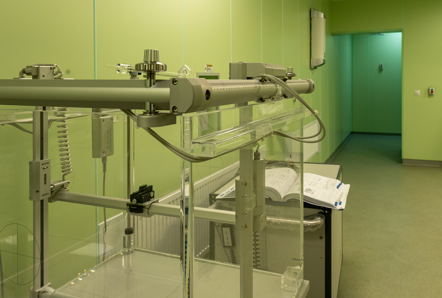
Large size motorized 3D water phantom system for dose distribution measurement of radiation therapy beams in real daily routine practice used as a part of quality control of radiation therapy.

What’s Inside Matters Most: Internal Dosimetry

Medical physics has been an integral part of medicine and healthcare over the greater part of the last century. Applying physics theory, concepts, and methods, scientists have created patient imaging, measurement, and treatment techniques that revolutionized the medical world. One product of medical physics has been the evolving specialty called radiopharmaceutical dosimetry, the calculation of absorbed dose and optimization of radiation dose delivery in cancer treatment. Today, we address internal dosimetry, the subset of medical physics that aims to optimize treatment and protect the patient from any undesirable side effects.

What is internal dosimetry?

Dosimetry is the measurement of radiation energy imparted to body organs and tissues. Radionuclides emit beneficial ionizing radiation that is useful for both diagnostic imaging of various diseases as well as for cancer treatment. Thus, medical internal dosimetry is the assessment of internal radiation dose from incorporated radionuclides associated with such life-saving radiopharmaceuticals.1 Radiation dose is the amount of energy imparted by radiations emitted during disintegration of radioactive atoms that constitute part of the radiopharmaceutical chemistry. Dose to organs of the body is quantified per unit mass (or weight) irradiated tissue. Dosimetry provides the fundamental quantities needed for several important purposes, including record-keeping, radiation protection decision-making, risk assessment, and cancer-treatment planning.2 The purpose and objective is to optimize medical benefit while minimizing potential radiation damage to body cells, tissues, and organs.

Dosimetry is a complex physical and biological science. Internal dosimetry provides critical information needed to better understand the biological mechanisms governing radionuclide uptake, translocation, and excretion from the body. The radiation dose imparted depends on the type, amount, and distribution of radionuclides, as well as specific nuclear properties, such as energy emitted.

Internal dosimetry differs from “external” dosimetry, which deals with the radiation dose from sources outside the body. Devices such as dosimeters measure external dosimetry directly, while internal dosimetry relies on indirect methods of radioactivity inside the patient using bioassay and imaging measurements.3 Bioassay is the measurement of the activity or concentration of radionuclides in biological samples. Samples can include urine specimens, feces, blood, or breath. Imaging techniques, such as whole-body counters or gamma cameras, can detect the radiation emitted by the radionuclides inside the body. This helps to provide information on their location and quantity.1

When is internal dosimetry used in healthcare?

Internal dosimetry mainly benefits patients who receive radionuclide therapy, a treatment that involves administering radioactively labeled proteins, such as monoclonal antibodies, to target specific types of cancer.3 It also helps to evaluate and account for unique patient variations in biodistribution—the way that different subjects respond to treatment. Internal dose assessments analyze radionuclide behavior in both normal (healthy) organs, as well as tumors. For example, imaging measurements provide physicists with important information to determine tumor uptake, retention, and clearance. In doing so, administered activity can be tailored according to patient health status, age, size, sex, and basal metabolic rates.

How does internal dosimetry produce useful data?

The main challenge of internal dosimetry is to track and follow the uptake, redistribution, metabolism, and clearance of the administered radiopharmaceutical inside the body over extended time periods after administration.4 Tracking sometimes involves mathematical modeling to describe the absorption, distribution, metabolization, and excretion of radionuclides by the body. Biokinetic models may be developed from the study of population groups, knowledge of radionuclide behavior in different organs, and the unique chemistry of each radiopharmaceutical. Biokinetic models incorporate mathematical compartments representing a particular organ or a tissue, and descriptions of the transfer rates that reflect the movement of radionuclides from one body compartment to another.1

Summary

Internal dosimetry is an important tool for radiation protection, especially in the fields of nuclear medicine, occupational health, and environmental monitoring. Dosimetry helps to customize or personalize nuclear medicine in cancer patients. In a broader sense, internal dosimetry is also applied to occupational and environmental health to prevent or reduce the exposure to radionuclides, by providing information on the sources, pathways, and levels of intake, and by suggesting appropriate measures, such as respiratory protection, contamination control, or dose limits. Internal dosimetry can also help to verify the adequacy of workplace controls, to demonstrate regulatory compliance, and to provide medical and legal evidence in case of accidental or intentional exposure.

Special software tools have been developed for the clinical nuclear medicine setting to facilitate medical imaging and calculate internal doses.

QDOSE® Multi-purpose Voxel Dosimetry (Personalized Dosimetry in Molecular Radiotherapy) is a complete, one-stop solution software for all internal dosimetry needs with multiple parallel workflows. With USFDA 510(k) clearance granted in August 2023, QDOSE® has proven its quality and compliance. To learn more, visit our QDOSE® webpage or schedule a meeting with our team.

Sources

  1. Sudprasert W, Belyakov OV, Tashiro S. Biological and internal dosimetry for radiation medicine: current status and future perspectives. J Radiat Res. 2022;63(2):247-254. doi:10.1093/jrr/rrab119
  2. Bartlett R, Bolch W, Brill AB, et al. MIRD Primer 2022: A Complete Guide to Radiopharmaceutical Dosimetry. Society of Nuclear Medicine & Molecular Imaging; 2022.
  3. Chapter 7 External and Internal Dosimetry. Accessed November 15, 2023. https://www.nrc.gov/docs/ML1121/ML11210B523.pdf
  4. What is Internal Dosimetry – Definition. Radiation Dosimetry. Published December 14, 2019. Accessed November 16, 2023. https://www.radiation-dosimetry.org/what-is-internal-dosimetry-definition/

13 Sep 2023
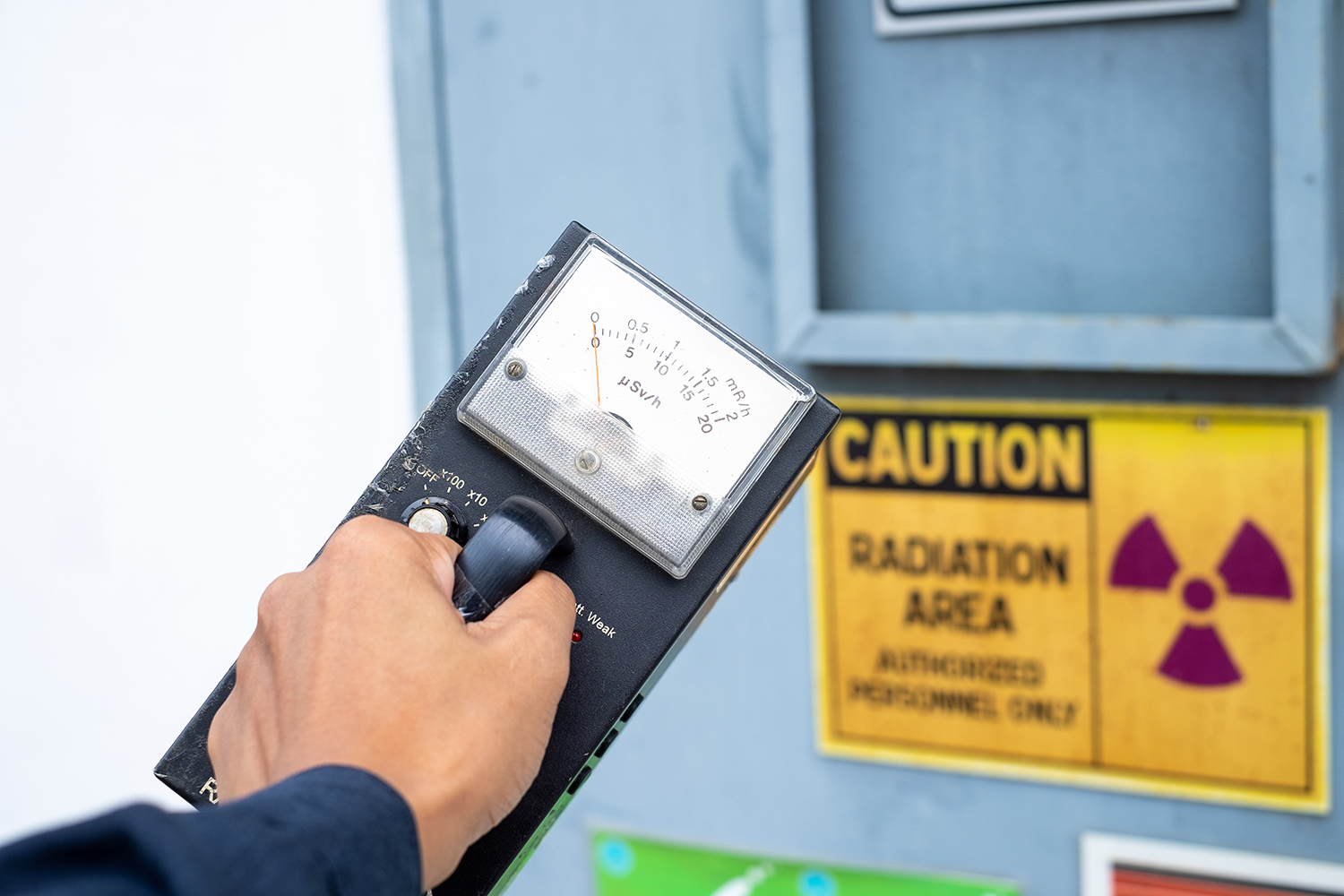
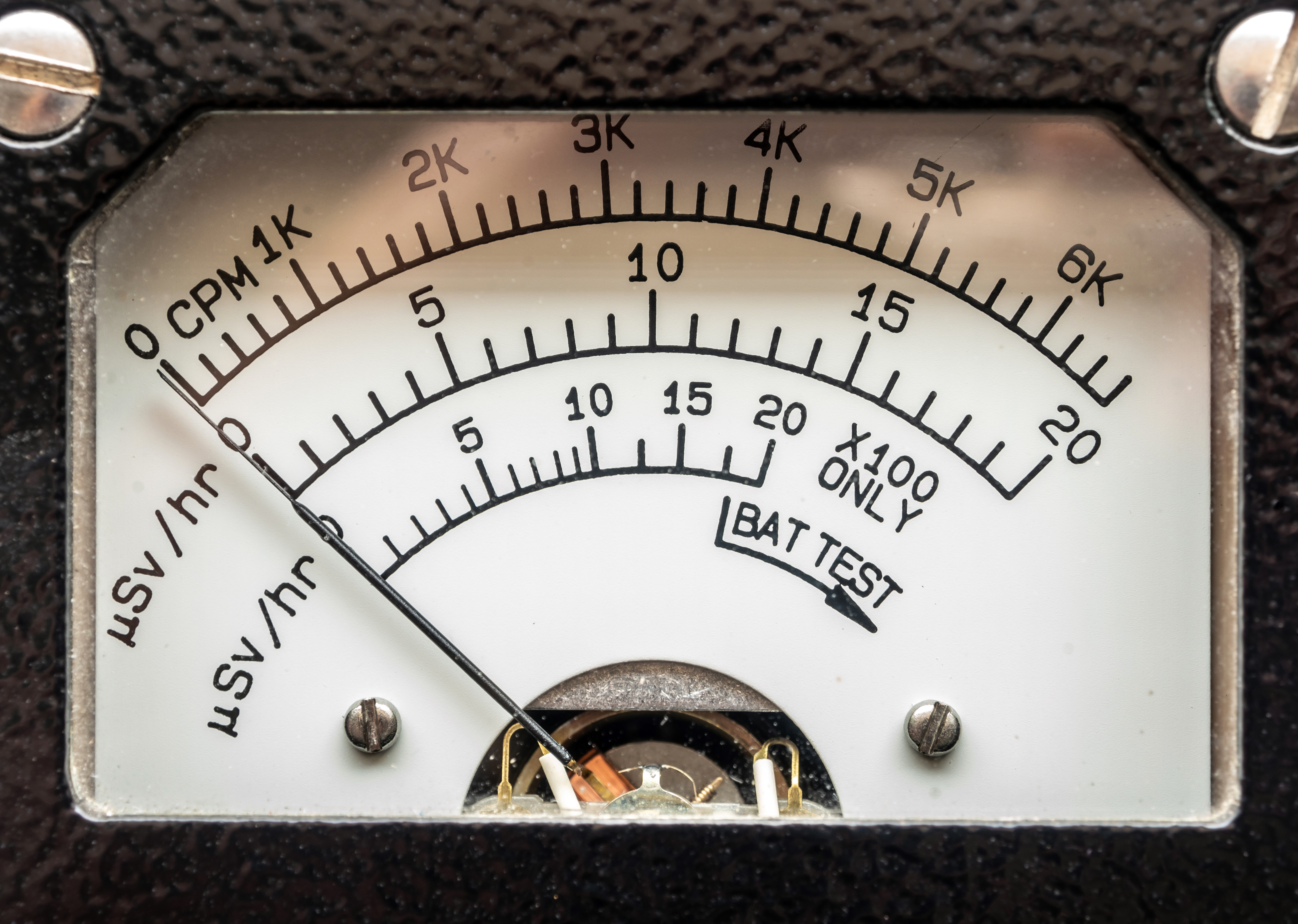
Supervisor use the survey meter to checks the level of radiation

The Units to Measure Radiation: Explained

The history of radiation units ties closely to the development of our understanding about radiation and its effects. The discovery of x-rays and radioactivity in the late 19th century by scientists like Wilhelm Roentgen, Henri Becquerel, and Pierre Curie paved the way for the exploration of radiation measurement.

As our knowledge of radiation’s effects on living organisms grew, the need for standardized units became evident. The roentgen was one of the earliest units to measure ionization, followed by the introduction of the curie to measure radioactivity. Over time, advancements in our understanding of radiation’s biological effects led to the development of units like the rem and the sievert.

Creating Radiation Units

The development of the SI system (International System of Units) established a standardized set of units to provide a coherent and universal way to measure radiation. The gray and the sievert were introduced as the primary units for absorbed dose and equivalent dose, respectively, within the SI system.

Four distinct yet interconnected units quantify radioactivity, exposure, absorbed dose, and dose equivalent. The mnemonic R-E-A-D creates a simple way to recall these units, which consist of a combination of commonly used (British, e.g., Ci) and internationally recognized (metric, e.g., Bq) units.1 Below, we will detail the mnemonic and discuss the history of the radiation units along with their relevant scientists:

Radioactivity defines the release of ionizing radiation from a substance. Whether it emits alpha or beta particles, gamma rays, x-rays, or neutrons, the radioactivity of a material is a measure of how many atoms within it decay over a specific period of time. The curie (Ci) and becquerel (Bq) units quantify radioactivity.1


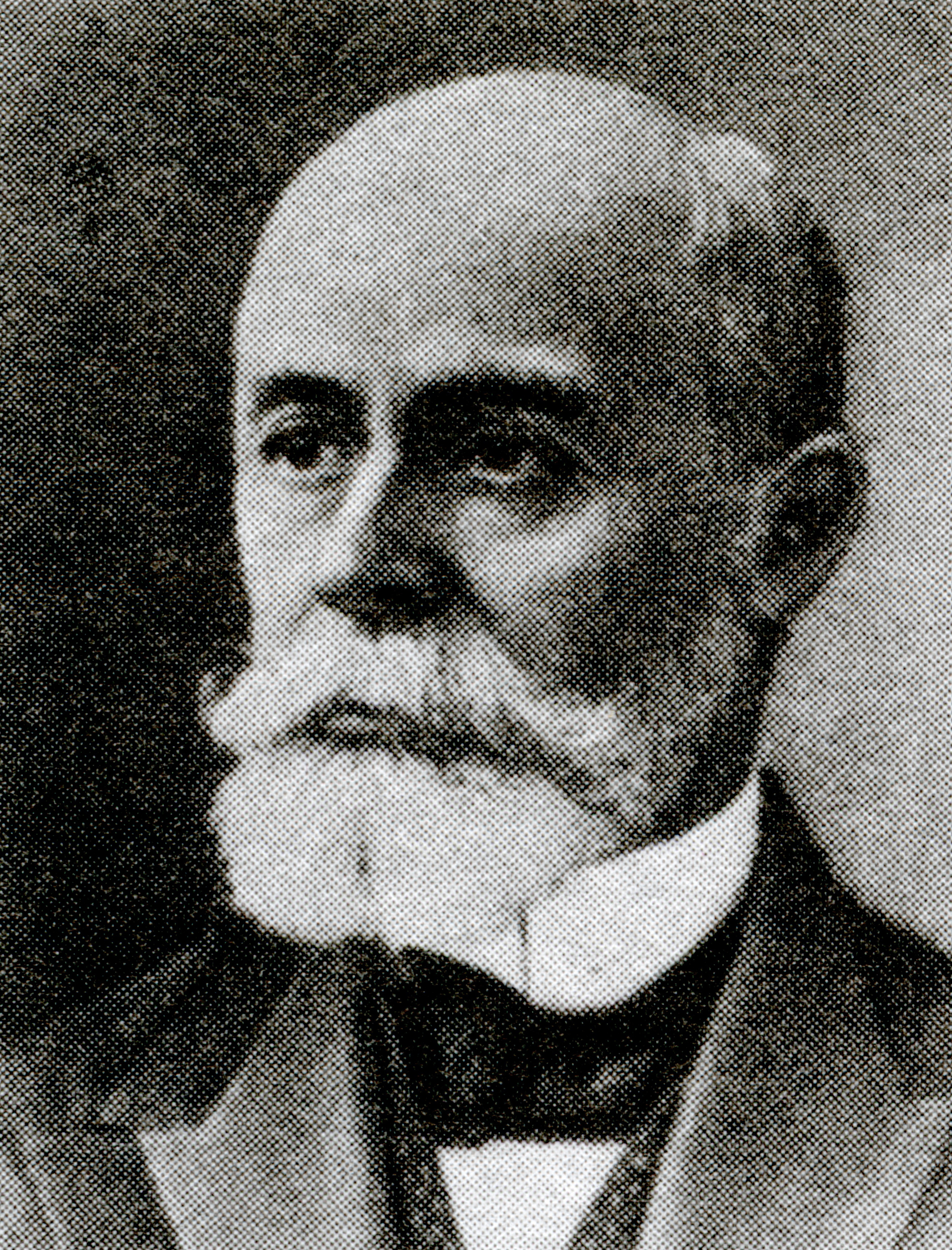
Antoine Henri Becquerel was a French physicist, engineer, and Nobel laureate who discovered evidence of radioactivity. Becquerel’s earliest works centered on the subject of his doctoral thesis: the plane polarization of light, with the phenomenon of phosphorescence and absorption of light by crystals. Early in his career, Becquerel also studied the Earth’s magnetic fields. In 1896, Becquerel discovered evidence of radioactivity while investigating phosphorescent materials such as some uranium salts. For his work in this field, he shared the 1903 Nobel Prize in Physics with Marie Curie and Pierre Curie. The SI unit for radioactivity, becquerel (Bq), is named after him.2

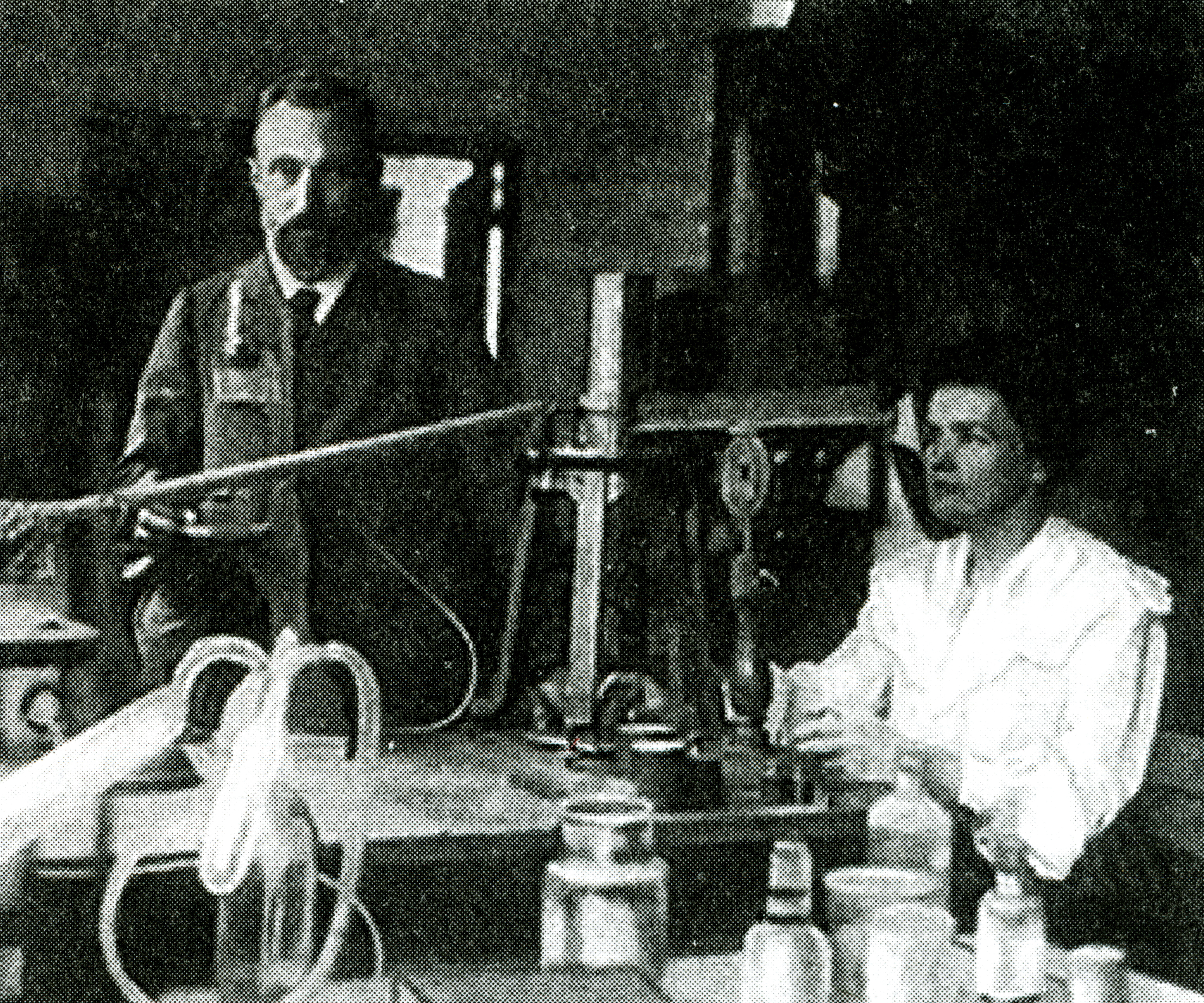
The curie (Ci) unit was created in 1910 by the International Congress of Radiology to measure radioactivity. Pierre Curie, another French physicist, and his wife Marie Curie, who also sat on the committee that named the unit, were the inspirations for the name through their radioactive studies. The original definition of the curie was “the quantity or mass of radium emanation in equilibrium with one gram of radium (element)”.3 In 1975, the becquerel replaced the curie as the official radiation unit in the International System of Units (SI) where 1 Bq = 1 nuclear decay/second4. The relationship between the two units is 1 Ci = 37 GBq (giga becquerels).

Exposure quantifies the extent of radiation going through the atmosphere that reaches a person’s body or a material. Numerous radiation monitors gauge exposure, utilizing the units of roentgen (R) or coulomb/kilogram (C/kg).1

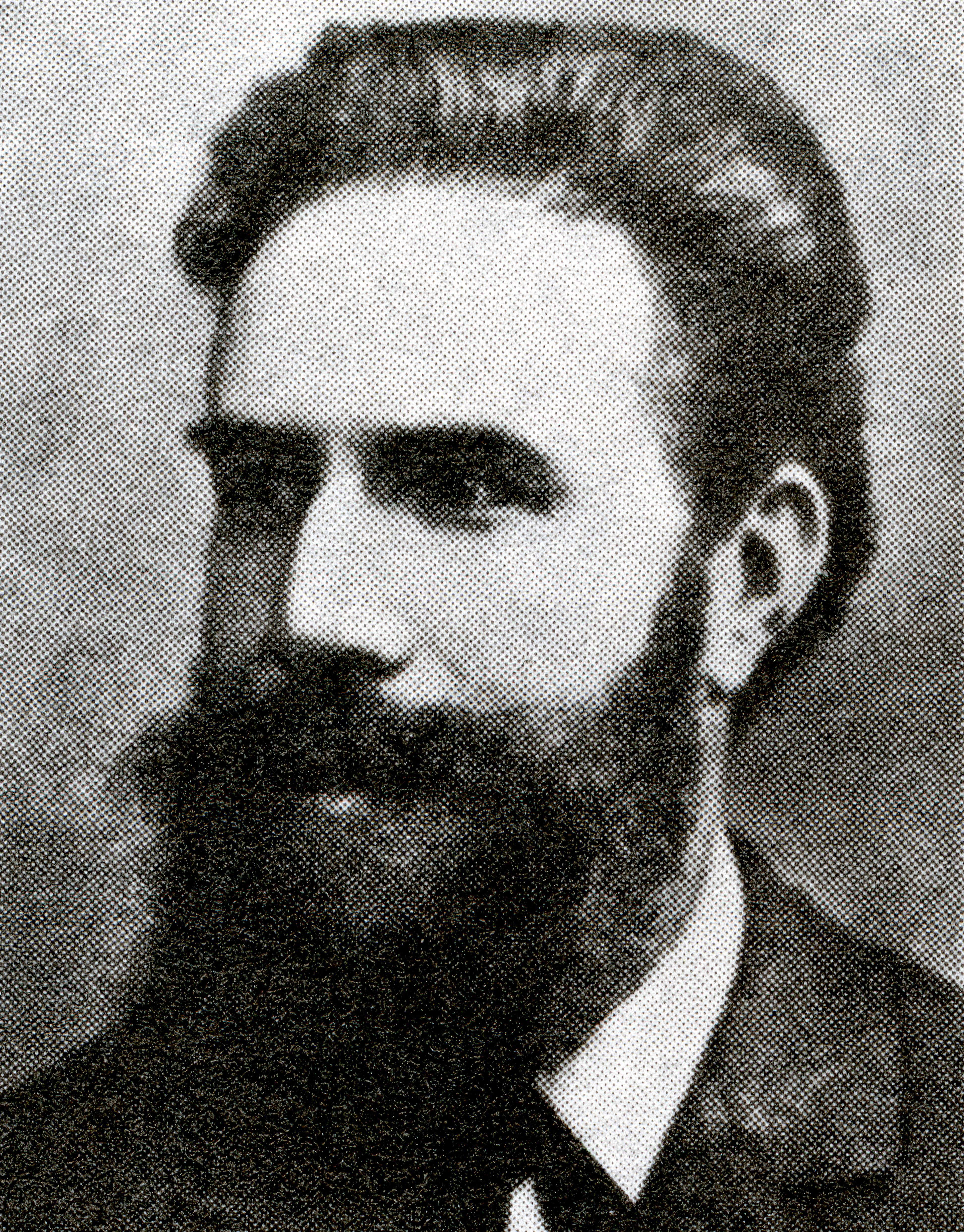
The roentgen is a legacy unit of measurement for the exposure of X-rays and gamma rays. This unit is defined as the electric charge freed by such radiation in a specified volume of air divided by the mass of that air and has the value 2.58 x 10⁻⁴ C/(kg air).5 It was named after Wilhelm Roentgen, a German physicist who discovered X-rays and was awarded the first Nobel Prize in Physics for the discovery.

In 1928, the roentgen became the first international measurement quantity for ionizing radiation defined for radiation protection. This is because it was, at the time, the most easily replicated method of measuring air ionization by using ion chambers.6 However, although this was a major step forward in standardizing radiation measurement, the roentgen had a disadvantage: it was only a measure of air ionization rather than a direct measure of radiation absorption in other materials, such as different forms of human tissue. As a result, it did not take into account the type of radiation or the biological effects of the different types of radiation on biological tissue. Consequently, new radiometric units for radiation protection came to be which took these concerns into account.7

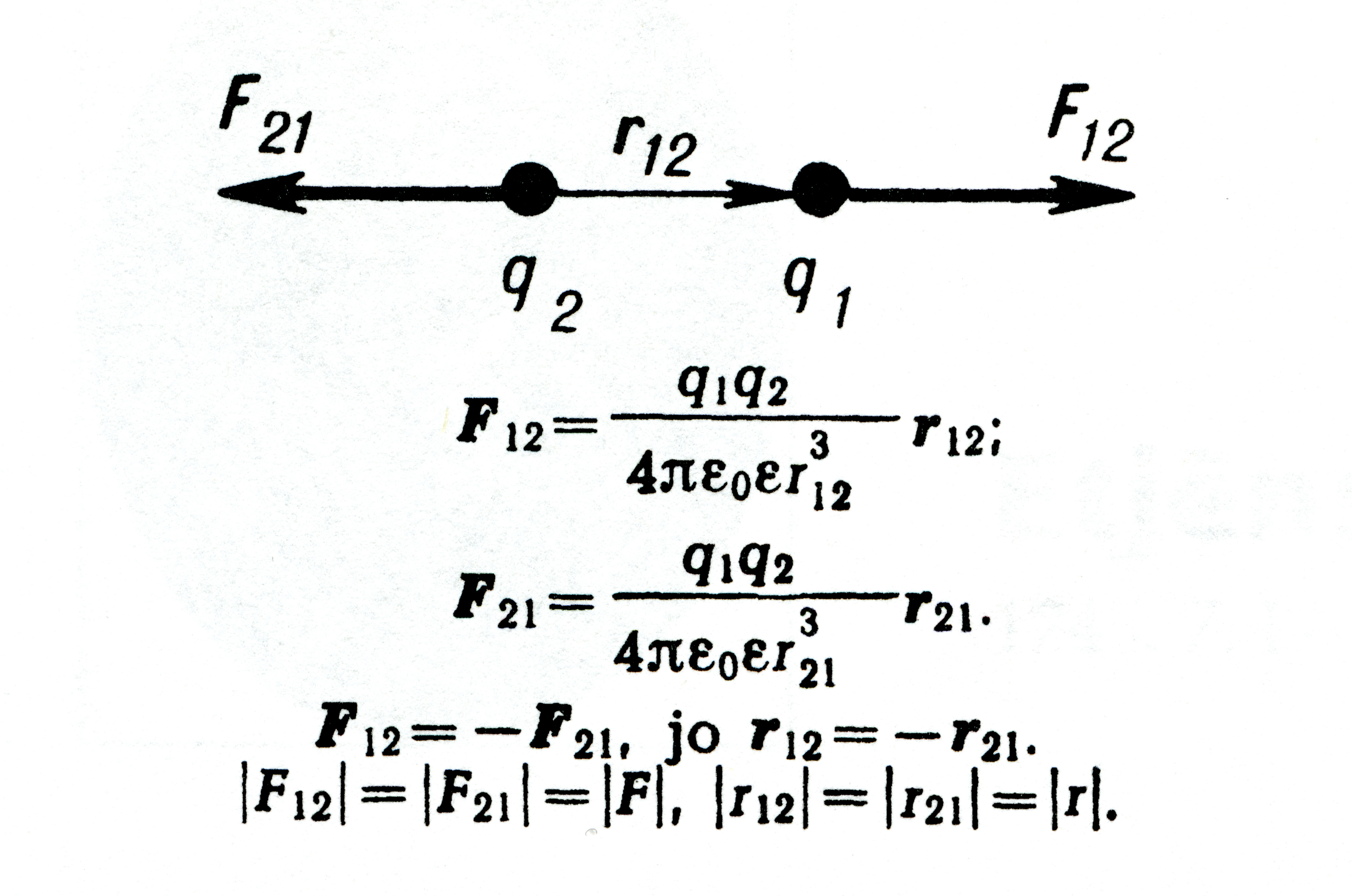
The SI unit for measuring exposure to ionizing radiation is coulomb per kilogram (C/kg). Interestingly, unlike other SI radiation units, this unit does not have a specific name. It officially replaced the previous unit, the roentgen, in 1975, with a transition period of at least ten years.8 The SI unit of electric charge, the coulomb, was named in honor of Charles-Augustin de Coulomb in 1880. Charles-Augustin de Coulomb was a French physicist whose best-known work is his formulation of Coulomb’s law. This law states that the force between two electrical charges is proportional to the product of the charges and inversely proportional to the square of the distance between them. He also made important contributions to the fields of electricity, magnetism, applied mechanics, friction studies, and torsion.9

Absorbed dose refers to the quantity of energy that is absorbed by an object or person where the energy is deposited by ionizing radiation as it passes through materials or the body. The units, radiation absorbed dose (rad) and gray (Gy), measure absorbed dose.1

In 1953, the International Commission on Radiation Units and Measurements (ICRU) adopted the unit rad at the Seventh International Congress of Radiology. This was the unit that replaced the rep, roentgen equivalent physical (detailed later in this blog). Although many believe that the rad is an abbreviation of “radiation absorbed dose”, the ICRU never identified it as such. This suggests that the term “rad” was as a standalone word to be a unit for absorbed dose. There was no documented discussion regarding the use of the rad prior to the Seventh International Congress of Radiology. The closest discussion was during the meeting in 1951 when they determined the need for this type of unit. In 1975, the gray (Gy) replaced the rad as the SI unit of absorbed dose where 1 Gy = 100 rad.10

Louis Harold Gray was a 20th century English physicist who worked mainly on the effects of radiation on biological systems. He was one of the earliest contributors to the field of radiobiology. He worked as a hospital physicist at Mount Vernon Hospital in London and developed the Bragg–Gray equation in collaboration with the father and son team of William Henry Bragg and William Lawrence Bragg. Bragg-Gray theory is the basis for the cavity ionization method of measuring energy absorption by materials exposed to ionizing radiation. Gray’s contributions to radiobiology were numerous. Amongst many other achievements, he developed the concept of RBE (Relative Biological Effectiveness) of doses of neutrons and initiated research into cells in hypoxic tumors and hyperbaric oxygen.11 Gray defined a unit of radiation dosage (absorbed dose) which was later named after him as an SI unit, the gray.

Dose equivalent, also known as effective dose, is a measurement that combines the amount of radiation absorbed and the impact it has on the human body. When it comes to beta and gamma radiation, the dose equivalent is equal to the absorbed dose. However, for alpha and neutron radiation, the dose equivalent surpasses the absorbed dose because these types of radiation have a greater biological impact resulting from their increased ability to damage tissue. To quantify dose equivalent, we use the units of roentgen equivalent man (rem) and sievert (Sv).1

Roentgen equivalent man, or “rem”, was first proposed for use in 1945, but under a different abbreviation. The roentgen was the only unit capable of expressing a radiation exposure at the time. However, it fell short being specifically measurable for photons. Workers came into contact with many other forms of radiation such as alpha particles, beta particles, and neutrons, so Herbert Parker, British-American physicist, created units that would be able to gauge exposures to many types of radiation. These were the roentgen equivalent physical (rep) and the roentgen equivalent biological (reb). Due to similarity in pronunciation between rep and reb, reb was eventually renamed to roentgen equivalent man or mammal (rem) to avoid confusion.

The first appearances of the rem unit in scientific literature were not until 1950.10 The rem related to the rad by multiplying the latter by a quality factor (QF) used to account for the varying biological effects of the different types of radiation. The rad in turn may be obtained from the roentgen by multiplying a dose conversion factor. In air, the dose conversion factor relationship between rad and roentgen is 1 R = 0.88 rad. The absorbed dose to a material is in turn found by multiplying 0.88 by the ratio of the mass energy absorption of the material to that of air.12

Rolf Maximilian Sievert was a Swedish medical physicist. He is best known for his work on the biological effects of ionizing radiation and his pioneering role in the measurement of doses of radiation, especially in its use in the diagnosis and treatment of cancer.13 Sievert contributed significantly to medical physics, earning him the title of “Father of Radiation Protection”. The sievert (Sv), the SI unit representing the stochastic health risk of ionizing radiation, is named after him. The sievert officially replaced the rem as the international SI unit in 1979 with 1 Sv = 100 rem.14

Conclusion

In summary, the history of radiation units is a journey that reflects the progress in our understanding of radiation’s properties and its impact on living organisms. The development of these units has enabled safer and more accurate measurement and assessment of radiation exposure and its effects on human health. By understanding the intricate relationship between radiation and our bodies, we can now take proactive measures to mitigate its harmful effects and promote a safer environment for all.

Sources

  1. NRC: Measuring Radiation. Nrc.gov. Published 2017. https://www.nrc.gov/about-nrc/radiation/health-effects/measuring-radiation.html
  2. The Nobel Prize. The Nobel Prize in Physics 1903. NobelPrize.org. Published 2019. https://www.nobelprize.org/prizes/physics/1903/becquerel/biographical/
  3. Curie – Unit of Radioactivity | nuclear-power.com. Nuclear Power. https://www.nuclear-power.com/nuclear-engineering/radiation-protection/units-of-radioactivity/curie-unit-of-radioactivity/
  4. ‌ Bell DJ. Becquerel (SI unit) | Radiology Reference Article | Radiopaedia.org. Radiopaedia. https://radiopaedia.org/articles/becquerel-si-unit
  5. ‌ Bashir U. Roentgen (unit) | Radiology Reference Article | Radiopaedia.org. Radiopaedia. Accessed August 29, 2023. https://radiopaedia.org/articles/roentgen-unit?lang=us
  6. ‌ Roentgen – Unit of Exposure | nuclear-power.com. Nuclear Power. Accessed August 29, 2023. https://www.nuclear-power.com/nuclear-engineering/radiation-protection/radiation-exposure/roentgen-unit-of-exposure/
  7. ‌ Roentgen (unit) explained. everything.explained.today. Accessed August 29, 2023. http://everything.explained.today/Roentgen_(unit)/
  8. ‌ Bell DJ. Coulomb per kilogram | Radiology Reference Article | Radiopaedia.org. Radiopaedia. Accessed August 29, 2023. https://radiopaedia.org/articles/coulomb-per-kilogram
  9. ‌ Laboratory NHMF. Charles-Augustin de Coulomb – Magnet Academy. nationalmaglab.org. https://nationalmaglab.org/magnet-academy/history-of-electricity-magnetism/pioneers/charles-augustin-de-coulomb/
  10. ‌ Why Did They Call It That? The Origin of Selected Radiological and Nuclear Terms. Museum of Radiation and Radioactivity. Accessed August 29, 2023. https://orau.org/health-physics-museum/articles/selected-radiological-nuclear-terms.html#rad
  11. ‌ LH Gray Memorial Trust: About L.H. Gray. www.lhgraytrust.org. Accessed August 29, 2023. http://www.lhgraytrust.org/lhgraybiography.html
  12. ‌Dosimetric Quantities and Units. U.S. NRC. Published October 25, 2010. Accessed September 11, 2023. https://www.nrc.gov/docs/ML1122/ML11229A688.pdf
  13. Aip.org. Published 2023. Accessed August 29, 2023. https://pubs.aip.org/physicstoday/Online/8433/Rolf-Sievert
  14. ‌ Bell DJ. Sievert (SI unit) | Radiology Reference Article | Radiopaedia.org. Radiopaedia. https://radiopaedia.org/articles/sievert-si-unit?lang=us