Tag: radiation safety

04 Nov 2025
A depiction of a nuclear power plant and its cooling towers.

 The Resurgence of Nuclear Energy

The world is in a new age of change and challenge with developments in technology, climate change, and available resources. Demands for further consideration on renewable energy options are rising, and with it comes a returning interest in nuclear energy. We can see the resurgence of nuclear energy in Versant Physics’ own home state: the Palisades Nuclear Power Plant, which stopped commercial operation in 2022, is being restored through a collaborative effort of Siemens Energy and Holtec International.1

These two companies aim to enhance safety, reliability, and efficiency through their efforts to modernize and renew the Palisades to generate more than 800 megawatts (MW) of electricity.2 The goal for this energy is to power around 800,000 households. Other major companies are pursuing similar restoration projects across the United States for different reasons. Let’s explore the history of nuclear energy, its fall out of favor, and why we’re seeing attention returning to the power source.

The Rise in Nuclear Reactor Development

The first technologies for nuclear reactors and creating nuclear energy came about in the 1930s and 1940s. Its potential as a clean energy source, however, was overshadowed by government demand for nuclear bomb research for World War II. It was not until the war came to an end that a renewed focus on nuclear energy could occur. The sheer capacity of nuclear power had been demonstrated in some of the most devastating ways, but a determination to harness it for good by way of creating steam and electricity rose in the 1950s.3

The first nuclear reactor to ever produce electricity was a small reactor created in the United States by Argonne National Laboratory in 1951. Seeing the rise in potential for power generation, President Eisenhower proposed an “Atoms for Peace” program in 1953. This reoriented research effort contributed significantly towards electricity generation and inspired civil nuclear energy development in the USA.  Great progress within the United States and the rest of the world continued throughout the 1950s with the creation of new reactor designs. These included fast breeder reactors (FBRs), which produce more fissile materials than they consume and are designed to extend nuclear fuel supply to generate electricity, as well as pressurized water reactors (PWRs), a reactor originally designed for the U.S. Navy that uses pressurized water as a coolant and neutron moderator.4,5

Commercial nuclear energy reactors started appearing in 1959 in France, quickly followed by the U.S., the United Kingdom, and Russia across the 1960s.  Many of these reactors were light water designs (either PWRs or boiling water reactors, BWRs) and some could generate up to 250 MW of energy. Within the early 1970s, the world saw its first high-power channel reactors that could generate 1,000 MW of energy.3 Despite these rapid developments of increasing energy outputs, the demand for this new source of power was short lived.

Why the Quick Decline to Nuclear Energy Interest?

Just as nuclear energy orders started coming in during the 1960s, the United States and other countries creating nuclear power plants began to see a decline just as quickly in the 1970s and through the rest of the 20th century.  This was due to unresolved concerns about the latest world war and unforeseen incidents in the future.

The Anti-Nuclear Movement and the Energy Crisis of the 1970s

 Within the midst of the Cold War where the world’s superpowers were locked in their nuclear arms race, animosity towards nuclear products spread throughout the public perspective. Local and national protests grew against the use of any nuclear weaponry or power plants due to the proven potential for destruction from World War II and the looming threat created by the Cold War.6 Producers of these plants were forced to bring production to a slowdown as previous orders from the 1960s were cancelled and interest in any new requests waned.3

In 1973 during the Yom Kippur War, the Arabian state members of the Organization of Petroleum Exporting Countries (OPEC) initiated an embargo on oil exports to the U.S. This created the oil crisis from 1973 which caused the U.S. government to scramble for alternative energy sources. This led to nuclear energy coming back on the table as an option for power, but concerns surrounding the consequences of power plants had only worsened. Within the U.S., many Americans were against storage of radioactive material and waste generated from the nuclear power plants. Protests cited potential danger to the health of residents living near the sites and the negative environmental impact if any waste leaked out of containment.6

The Chernobyl, Three Miles Island, and The Fukushima Disaster Incidents

After the rise of the Anti-Nuclear Movement, incidents relating to established nuclear power plants began to occur. The first recorded nuclear power plant incident happened in 1979 at the Three Mile Island nuclear power plant in the USA. Due to a cooling malfunction, part of the core in Unit 2 of the plant melted, destroying the reactor. Although there were no injuries or adverse health effects from the event, the event caused widespread confusion and concern about the impact of the incident.7

The Chernobyl disaster occurred in 1986 and is notably the worst incident to have occurred in history. This nuclear power plant is in Chernobyl, Ukraine, and a sudden surge of power during a reactor systems test destroyed Unit 4 of the station. Investigation of the incident determined that a lack of proper safety culture caused the devastation at the Chernobyl power plant8. Many site workers were killed or had acute radiation sickness.9

Image depicting a full view of the Chernobyl nuclear power plant.

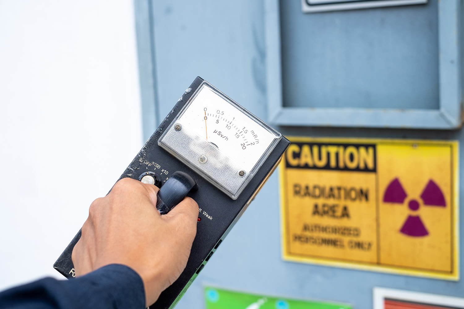
These incidents only heightened anti-nuclear movements in the end of the 20th century. Due to the strong reluctance towards increasing nuclear energy production, new facilities were rare into the early 2000s.  The Fukushima Daiichi accident of 2011, in which an unprecedented natural disaster hit Japan and overpowered the outdated tsunami countermeasures of the Daiichi site, continued to incite trepidation towards the safety of nuclear power plants.10 The world is seeing more alternative energy sources in response to climate change, but through solar, wind, and geothermal routes. Nuclear energy produces only 9% of the world’s electricity as of 2025,11 but things may change soon. After over a decade without serious troubles, eyes are returning to nuclear energy as power for new technologies.

Nuclear Energy’s New Appeal

One of the biggest technological advancements that the world is adapting to is the rise in artificial intelligence (AI). Companies across the globe are implementing their own AI systems that require very large, energy demanding data centers to function. For one request to ChatGPT, the International Energy Agency (IEA) has determined that the response the AI provides requires ten times more electricity than someone completing a Google search.12 Current power grids are struggling to keep up with the demands of artificial intelligence. In response, companies creating AI who need to maintain these data centers are casting attention towards untapped nuclear energy sources.

Who are some of the big names looking to harness nuclear energy?

One of the first deals made that suggested a reemergence of nuclear energy was in September of 2024. The tech giant Microsoft signed an agreement with Constellation Energy to restart the Unit 1 nuclear reactor at the Three Mile Island plant in Pennsylvania.13 Once the site is up and running, Microsoft hopes to have 835 megawatts (MW) of new power they can dedicate to their data centers. Whether this $1.6 billion deal is worth its steep price tag depends on time—the Three Mile Island plant originally closed in 2019 due to economic challenges. Now, Microsoft faces getting the nuclear reactors back into working order, renewing its operating licenses, and starting operations again by 2028.13

The social media and technology giant, Meta, aims to take a similar approach to Microsoft for their AI power needs. By partnering with Constellation Energy, Meta hopes to access 1,121 MW of nuclear energy through an established Illinois nuclear facility. This will bolster the electricity needs for Meta to continue its AI developments. They hope to increase data centers and important hubs for tasks like “AI model training, content delivery, and cloud services”.14

While Microsoft and Meta will be taking advantage of established nuclear power plants, Google and Amazon are approaching their move into nuclear energy differently. These companies are looking to invest in the creation of small modular reactors (SMRs). This type of plant currently doesn’t operate anywhere in the United States and only a few exist in the world. SMRs produce less than 300 MW per reactor. Amazon has made a deal with X-Energy, a company capable of designing “high-temperature gas reactors,” and Energy Northwest. They aim to create four reactor modules that would provide at least 320 MW combined.14 Meanwhile, Google has moved into an agreement with Kairos, a company that develops “molten salt-cooled, TRISO fuel-powered” reactors. The goal of power production from these future SMRs for Google would be 500 MW by 2035.14

Will nuclear energy be the long-term solution for AI needs?

The unexpected energy demand for artificial intelligence is driving these trillion-dollar companies to invest in nuclear energy potential, but will this be a long-term investment? Some suggest that the eventual solution to this power-hungry technology will be the technology itself. Artificial intelligence is continuously growing, being trained to learn more and do more. Some believe that AI algorithms will eventually become the best path to determining its own energy management. This would be achievable through its capacity to “identify patterns in data, detect anomalies, and anticipate and forecast future results”.12

Given enough time to “self-reflect”, AI could determine how to optimize its own functioning to reduce its carbon footprint and power necessary to be sustained. In the meantime, nuclear power plants will be giving these tech giants an opportunity to obtain the energy they need. With the increasing pressure of honoring climate commitments and lowering greenhouse gases, they may also find that nuclear energy suits more than just their AI needs.

Conclusion

Nuclear energy is still an industry that is seen first for the disastrous events from the past and their repercussions. Power plants are considered as hazardous potential for nuclear waste leaks, core meltdowns, and a threat for radiation exposure to those who live nearby. However, as the need for cleaner energy increases in demand, we may find that nuclear is an option to reconsider.

Handling of radiation and nuclear power has improved over recent decades. Now more than ever, safety concerns are taken seriously to avoid repetitions of historical incidents. With investments from large companies like Microsoft and Meta, the resurgence of nuclear energy could provide a path to expanded opportunities of clean power provision for more than just AI data centers. By focusing on safety, responsible handling, and sustainability as core features of future nuclear power plants, perhaps the true potential for nuclear energy will be revealed.

Sources

  1. Palisades Nuclear Power Plant. Siemens-energy.com. Published 2025. Accessed October 30, 2025. https://www.siemens-energy.com/global/en/home/references/recommissioning-palisades-nuclear.html
  2. Davidson K. Palisades Nuclear Plant on Lake Michigan shoreline one step closer to reopening • Michigan Advance. Michigan Advance. Published July 25, 2025. Accessed October 30, 2025. https://michiganadvance.com/briefs/palisades-nuclear-plant-on-lake-michigan-shoreline-one-step-closer-to-reopening/
  3. ‌World Nuclear Association. Outline History of Nuclear Energy. world-nuclear.org. Published May 2, 2024. https://world-nuclear.org/information-library/current-and-future-generation/outline-history-of-nuclear-energy
  4. Breeder reactor – Energy Education. Energyeducation.ca. Published 2017. https://energyeducation.ca/encyclopedia/Breeder_reactor
  5. World Nuclear Association. Nuclear Power Reactors. world-nuclear.org. Published January 23, 2025. https://world-nuclear.org/information-library/nuclear-fuel-cycle/nuclear-power-reactors/nuclear-power-reactors
  6. Antinuclear movement of the 1970s | EBSCO. EBSCO Information Services, Inc. | www.ebsco.com. Published 2022. https://www.ebsco.com/research-starters/history/antinuclear-movement-1970s
  7. World Nuclear Association. Three Mile Island Accident – World Nuclear Association. world nuclear association. Published October 11, 2022. https://world-nuclear.org/information-library/safety-and-security/safety-of-plants/three-mile-island-accident
  8. World Nuclear Association. Sequence of Events – Chernobyl Accident Appendix 1 – World Nuclear Association. World-nuclear.org. Published 2022. https://world-nuclear.org/information-library/appendices/chernobyl-accident-appendix-1-sequence-of-events
  9. Backgrounder on Chernobyl Nuclear Power Plant Accident. NRC Web. Published 2015. https://www.nrc.gov/reading-rm/doc-collections/fact-sheets/chernobyl-bg
  10. World Nuclear Association. Fukushima Daiichi Accident. world-nuclear.org. Published April 29, 2024. https://world-nuclear.org/information-library/safety-and-security/safety-of-plants/fukushima-daiichi-accident
  11. Nuclear Power Reactors – World Nuclear Association. world-nuclear.org. https://world-nuclear.org/information-library/nuclear-fuel-cycle/nuclear-power-reactors/nuclear-power-reactors#how-does-a-nuclear-reactor-work
  12. United Nations. Artificial intelligence: How much energy does AI use? United Nations Western Europe. Published April 7, 2025. https://unric.org/en/artificial-intelligence-how-much-energy-does-ai-use/
  13. Moseman A. How to Restart a Nuclear Reactor. IEEE Spectrum. Published October 2, 2024. https://spectrum.ieee.org/three-mile-island
  14. Wolinski SJ. AI’s Energy Demands and Nuclear’s Uncertain Future | GJIA. Georgetown Journal of International Affairs. Published April 16, 2025. https://gjia.georgetown.edu/2025/04/16/ais-energy-demands-and-nuclears-uncertain-future/

27 Mar 2024
Airplane flight crew character design. Pilot and stewardess flat vector illustration

Flight Crews and Radiation Exposure

Flight crews are among the occupational groups most exposed to ionizing radiation, with an average annual effective dose surpassing that of other radiation-exposed workers in the United States, excluding astronauts.1 This elevated exposure is primarily due to the high levels of cosmic radiation encountered at flight altitudes, which can pose significant health risks to pilots and cabin crew members.2 In this blog post, we’ll explore the nature of cosmic radiation, its potential health effects, current exposure levels for aircrews, as well as the guidelines and regulations in place to ensure their safety.

What Is Cosmic Ionizing Radiation?

As we’ve touched on in a previous blog, cosmic ionizing radiation–or simply cosmic radiation–originates from beyond Earth’s atmosphere. Additionally, it consists of two main components: galactic cosmic radiation (GCR) and solar particle events (SPEs).3,4

Galactic Cosmic Radiation

GCR is a constant background radiation that permeates interstellar space, originating from distant stars and galaxies. It is composed primarily of high-energy protons (85%) and alpha particles (14%). There is also a small fraction of heavier nuclei (1%) ranging from lithium to iron and beyond. These particles span a wide energy range, and as a result some reach extremely high energies capable of penetrating deep into the Earth’s atmosphere and passing through aircraft shielding.5

Solar Particle Events (Solar Flares)

Solar particle events, on the other hand, are sporadic bursts of intense radiation associated with solar flares and coronal mass ejections. During an SPE, the Sun ejects a large number of high-energy protons and other particles that can reach Earth within hours to days. While less frequent than GCR, SPEs can dramatically increase radiation exposure for flight crews, particularly those on polar routes where the Earth’s magnetic field provides less protection.6,7

At higher altitudes, such as those typically encountered during air travel, the Earth’s atmosphere provides less shielding against cosmic radiation, resulting in increased exposure for flight crews and passengers.

Several studies that have investigated the difference in cosmic ray levels at various altitudes versus ground level found that the dose rate of cosmic radiation at a cruising altitude of 30,000 feet was approximately 10 times higher than at sea level.

The specific increase in cosmic ray exposure at higher altitudes is influenced by several factors, including the solar cycle (solar maximum vs. solar minimum), geomagnetic field strength, and also the path of the flight (polar routes are exposed to higher levels of cosmic rays). For example, during periods of high solar activity (solar maximum), the increased solar wind can actually shield the Earth from some cosmic rays, slightly reducing the exposure at high altitudes. Conversely, during a solar minimum, the cosmic ray intensity can be higher.

Estimates of the number of hours that have to be flown in order to receive an effective dose of 1 mSv at 30,000 feet are 510 hours at a latitude of 30o South and 1,330 hours at the equator.8

Health Effects and Uncertainties

The health risks associated with radiation exposure are generally well-documented. Prolonged exposure to high levels of radiation can increase the risk of cancer, cataracts, as well as other adverse health effects.  However, quantifying the specific risks associated with the chronic low-dose radiation experienced by flight crews remains a challenge.

The World Health Organization’s International Agency for Research on Cancer (IARC) acknowledges that ionizing radiation causes cancer in humans and is also associated with reproductive problems. However, when it comes to cosmic ionizing radiation, several uncertainties remain:

  1. Cancer Risk: Most radiation health studies have focused on groups exposed to much higher doses from different types of radiation (such as atomic bomb survivors or patients receiving radiation therapy).9 Due to this, the specific link between cosmic ionizing radiation and cancer risk is not yet fully understood.
  2. Reproductive Health: Miscarriages and birth defects related to cosmic radiation exposure are still not definitively established.10

Despite the limitations of current research, several studies have suggested that flight crews may have a higher incidence of certain cancers compared to the general population. These include breast cancer, melanoma, as well as non-melanoma skin cancers.11,12 However, the causal link between cosmic radiation exposure and these increased risks has not been definitively established. Other factors, such as lifestyle and genetic predisposition, may also play a role.13

Exposure Levels for Flight Crews

Recent dose and risk assessments by a wide variety of investigators have demonstrated the need to dedicate further attempts to quantify potential radiation exposure.14 The National Council on Radiation Protection and Measurements (NCRP) reports an average annual effective dose of 3.07 mSv for flight crews; most of this exposure comes from natural radiation:

  • Estimates of annual aircrew cosmic radiation exposure range from 0.2 to 5 millisieverts (mSv) per year depending on factors such as flight routes, altitude, and solar activity.
  • Solar particle events occur less frequently, but during events, exposure levels can increase substantially and potentially lead to higher doses over short periods.

Guidelines and Regulations

While there are no official dose limits specifically for aircrew in the United States, national and international guidelines provide context:

  • International Commission on Radiological Protection (ICRP): Recognizes aircrew as radiation-exposed workers. They also recommend an effective dose limit of 20 mSv per year averaged over 5 years (totaling 100 mSv in 5 years) for radiation workers. However, for the general public, the recommended limit is 1 mSv per year.15
  • Pregnant Aircrew: The ICRP recommends a dose limit of 1 mSv throughout pregnancy.16

Current regulations aim to limit radiation exposure for flight crews, but there is room for improvement. The International Commission on Radiological Protection (ICRP) sets guidelines for radiation protection and also includes dose limits for occupational exposure. However, these guidelines may not adequately address the unique challenges faced by flight crews. To improve current radiation safety regulations for aircrews, a multi-faceted approach is necessary. This should include:

Improved Monitoring and Data Collection

Implementing advanced radiation monitoring systems on aircraft in addition to encouraging the use of personal dosimeters by flight crews can provide more accurate and comprehensive data on exposure levels17. This information can help refine risk assessments as well as guide the development of more effective protection strategies.

Aircraft Shielding and Design

Continued research into advanced shielding materials in addition to aircraft design modifications can help reduce the radiation dose received by flight crews and passengers18. This may also involve the use of novel composite materials or the incorporation of additional shielding in critical areas of the aircraft.

Route Optimization and Flight Planning

By carefully planning flight routes and altitudes, airlines can minimize exposure to cosmic radiation, particularly during solar particle events19. This may also involve rerouting flights to lower latitudes or reducing flight time at higher altitudes when necessary.

Education and Awareness Programs

Providing flight crews with comprehensive information about the risks of cosmic radiation exposure in addition to the importance of proper protection measures can empower them to make informed decisions about their health and safety20. This should include training on the use of personal protective equipment, such as dosimeters, as well as guidelines for managing exposure during pregnancy.

Regulatory Harmonization and Enforcement

Strengthening international collaboration to harmonize radiation protection standards for flight crews in addition to ensuring consistent implementation and enforcement of these standards across the aviation industry can help create a safer working environment for all aircrews21.

Conclusion

Although no regulations officially set dose limits, radiation exposure is still a concern to be evaluated for airplane flight crews due to their occupational exposure to cosmic radiation. While the specific health risks associated with this chronic low-dose exposure remain uncertain, continued efforts are essential ensure a safe working environment. By implementing measures such as personal dosimetry devices, increased monitoring, staff training, and encouraging airplane manufacturers to consider shielding and design modifications, airlines can better protect their flight crews. Ensuring a safer career for every radiation worker will require time, dedication, and collaboration. However, the benefits for the health and safety of all industries, including aircrews, make it a worthwhile endeavor.

Versant Physics is a full-service medical physics and radiation safety consulting company based in Kalamazoo, MI. Contact us for all of your regulatory, radiation safety, and personnel dosimetry needs.

Sources

  1. Friedberg, W., & Copeland, K. (2003). What aircrews should know about their occupational exposure to ionizing radiation. Oklahoma City, OK: Civil Aerospace Medical Institute, Federal Aviation Administration. ↩︎
  2. United Nations Scientific Committee on the Effects of Atomic Radiation. (2008). Sources and effects of ionizing radiation: UNSCEAR 2008 report to the General Assembly, with scientific annexes. New York: United Nations. ↩︎
  3. Validation of modelling the radiation exposure due to solar particle events at aircraft altitudes. Radiation Protection Dosimetry, Volume 131, Issue 1, August 2008, Pages 51–58. https://doi.org/10.1093/rpd/ncn238 ↩︎
  4. Wilson, J. W., Townsend, L. W., Schimmerling, W., Khandelwal, G. S., Khan, F., Nealy, J. E.,  & Norbury, J. W. (1991). Transport methods and interactions for space radiations. NASA Reference Publication, 1257 ↩︎
  5. O’Sullivan, D. Exposure to galactic cosmic radiation and solar energetic particles. Radiat Prot Dosimetry. 2007;125(1-4):407-11. https://pubmed.ncbi.nlm.nih.gov/17846031/ ↩︎
  6. Turner, R. E. (2007). Solar particle events from a risk management perspective. Radiation Protection Dosimetry, 127(1-4), 534-538. ↩︎
  7. Lantos, P., & Fuller, N. (2003). History of the solar particle event radiation doses on-board aeroplanes using a semi-empirical model and Concorde measurements. Radiation Protection Dosimetry, 104(3), 199-210. ↩︎
  8. Cosmic Radiation Exposure for Casual Flyers and Aircrew, https://www.arpansa.gov.au/understanding-radiation/radiation-sources/more-radiation-sources/flying-and-health ↩︎
  9. National Research Council. (2006). Health risks from exposure to low levels of ionizing radiation: BEIR VII phase 2 (Vol. 7). National Academies Press. ↩︎
  10. CDC – Aircrew Safety and Health – Cosmic Ionizing Radiation – NIOSH Workplace Safety & Health Topics. Centers for Disease Control and Prevention. Published 2019. https://www.cdc.gov/niosh/topics/aircrew/cosmicionizingradiation.html ↩︎
  11. Pukkala, E., Aspholm, R., Auvinen, A., Eliasch, H., Gundestrup, M., Haldorsen, T., & Tveten, U. (2003). Cancer incidence among 10,211 airline pilots: a Nordic study. Aviation, Space, and Environmental Medicine, 74(7), 699-706. ↩︎
  12. Rafnsson, V., Hrafnkelsson, J., & Tulinius, H. (2000). Incidence of cancer among commercial airline pilots. Occupational and Environmental Medicine, 57(3), 175-179. ↩︎
  13. Hammer, G. P., Blettner, M., & Zeeb, H. (2009). Epidemiological studies of cancer in aircrew. Radiation Protection Dosimetry, 136(4), 232-239. ↩︎
  14. Olumuyiwa A. Occupational Radiation Exposures in Aviation: Air Traffic Safety Systems Considerations. International Journal of Aviation, Aeronautics, and Aerospace. Published online 2020. doi:https://doi.org/10.15394/ijaaa.2020.1476 ↩︎
  15. International Commission on Radiological Protection. (2007). The 2007 recommendations of the International Commission on Radiological Protection. Annals of the ICRP, 37(2-4), 1-332. ↩︎
  16. International Commission on Radiological Protection. (2000). Pregnancy and medical radiation. Annals of the ICRP, 30(1), iii-viii, 1-43. ↩︎
  17. Bartlett, D. T. (2004). Radiation protection aspects of the cosmic radiation exposure of aircraft crew. Radiation Protection Dosimetry, 109(4), 349-355. ↩︎
  18. Wilson, J. W., Miller, J., Konradi, A., & Cucinotta, F. A. (1997). Shielding strategies for human space exploration. NASA Conference Publication, 3360. ↩︎
  19. Copeland, K. (2014). Cosmic radiation and commercial air travel. Radiation Protection Dosimetry, 162(3), 351-357. ↩︎
  20. International Civil Aviation Organization. (2012). Manual of Civil Aviation Medicine. https://www.icao.int/publications/Documents/8984_cons_en.pdf ↩︎
  21. International Atomic Energy Agency: Cosmic radiation exposure of aircrew and space crew. https://www.iaea.org/sites/default/files/20/11/rasa-cosmic.pdf ↩︎
13 Sep 2023
Supervisor use the survey meter to checks the level of radiation

The Units to Measure Radiation: Explained

The history of radiation units ties closely to the development of our understanding about radiation and its effects. The discovery of x-rays and radioactivity in the late 19th century by scientists like Wilhelm Roentgen, Henri Becquerel, and Pierre Curie paved the way for the exploration of radiation measurement.

As our knowledge of radiation’s effects on living organisms grew, the need for standardized units became evident. The roentgen was one of the earliest units to measure ionization, followed by the introduction of the curie to measure radioactivity. Over time, advancements in our understanding of radiation’s biological effects led to the development of units like the rem and the sievert.

Creating Radiation Units

The development of the SI system (International System of Units) established a standardized set of units to provide a coherent and universal way to measure radiation. The gray and the sievert were introduced as the primary units for absorbed dose and equivalent dose, respectively, within the SI system.

Four distinct yet interconnected units quantify radioactivity, exposure, absorbed dose, and dose equivalent. The mnemonic R-E-A-D creates a simple way to recall these units, which consist of a combination of commonly used (British, e.g., Ci) and internationally recognized (metric, e.g., Bq) units.1 Below, we will detail the mnemonic and discuss the history of the radiation units along with their relevant scientists:

Radioactivity defines the release of ionizing radiation from a substance. Whether it emits alpha or beta particles, gamma rays, x-rays, or neutrons, the radioactivity of a material is a measure of how many atoms within it decay over a specific period of time. The curie (Ci) and becquerel (Bq) units quantify radioactivity.1


Antoine Henri Becquerel was a French physicist, engineer, and Nobel laureate who discovered evidence of radioactivity. Becquerel’s earliest works centered on the subject of his doctoral thesis: the plane polarization of light, with the phenomenon of phosphorescence and absorption of light by crystals. Early in his career, Becquerel also studied the Earth’s magnetic fields. In 1896, Becquerel discovered evidence of radioactivity while investigating phosphorescent materials such as some uranium salts. For his work in this field, he shared the 1903 Nobel Prize in Physics with Marie Curie and Pierre Curie. The SI unit for radioactivity, becquerel (Bq), is named after him.2

The curie (Ci) unit was created in 1910 by the International Congress of Radiology to measure radioactivity. Pierre Curie, another French physicist, and his wife Marie Curie, who also sat on the committee that named the unit, were the inspirations for the name through their radioactive studies. The original definition of the curie was “the quantity or mass of radium emanation in equilibrium with one gram of radium (element)”.3 In 1975, the becquerel replaced the curie as the official radiation unit in the International System of Units (SI) where 1 Bq = 1 nuclear decay/second4. The relationship between the two units is 1 Ci = 37 GBq (giga becquerels).

Exposure quantifies the extent of radiation going through the atmosphere that reaches a person’s body or a material. Numerous radiation monitors gauge exposure, utilizing the units of roentgen (R) or coulomb/kilogram (C/kg).1

The roentgen is a legacy unit of measurement for the exposure of X-rays and gamma rays. This unit is defined as the electric charge freed by such radiation in a specified volume of air divided by the mass of that air and has the value 2.58 x 10⁻⁴ C/(kg air).5 It was named after Wilhelm Roentgen, a German physicist who discovered X-rays and was awarded the first Nobel Prize in Physics for the discovery.

In 1928, the roentgen became the first international measurement quantity for ionizing radiation defined for radiation protection. This is because it was, at the time, the most easily replicated method of measuring air ionization by using ion chambers.6 However, although this was a major step forward in standardizing radiation measurement, the roentgen had a disadvantage: it was only a measure of air ionization rather than a direct measure of radiation absorption in other materials, such as different forms of human tissue. As a result, it did not take into account the type of radiation or the biological effects of the different types of radiation on biological tissue. Consequently, new radiometric units for radiation protection came to be which took these concerns into account.7

The SI unit for measuring exposure to ionizing radiation is coulomb per kilogram (C/kg). Interestingly, unlike other SI radiation units, this unit does not have a specific name. It officially replaced the previous unit, the roentgen, in 1975, with a transition period of at least ten years.8 The SI unit of electric charge, the coulomb, was named in honor of Charles-Augustin de Coulomb in 1880. Charles-Augustin de Coulomb was a French physicist whose best-known work is his formulation of Coulomb’s law. This law states that the force between two electrical charges is proportional to the product of the charges and inversely proportional to the square of the distance between them. He also made important contributions to the fields of electricity, magnetism, applied mechanics, friction studies, and torsion.9

Absorbed dose refers to the quantity of energy that is absorbed by an object or person where the energy is deposited by ionizing radiation as it passes through materials or the body. The units, radiation absorbed dose (rad) and gray (Gy), measure absorbed dose.1

In 1953, the International Commission on Radiation Units and Measurements (ICRU) adopted the unit rad at the Seventh International Congress of Radiology. This was the unit that replaced the rep, roentgen equivalent physical (detailed later in this blog). Although many believe that the rad is an abbreviation of “radiation absorbed dose”, the ICRU never identified it as such. This suggests that the term “rad” was as a standalone word to be a unit for absorbed dose. There was no documented discussion regarding the use of the rad prior to the Seventh International Congress of Radiology. The closest discussion was during the meeting in 1951 when they determined the need for this type of unit. In 1975, the gray (Gy) replaced the rad as the SI unit of absorbed dose where 1 Gy = 100 rad.10

Louis Harold Gray was a 20th century English physicist who worked mainly on the effects of radiation on biological systems. He was one of the earliest contributors to the field of radiobiology. He worked as a hospital physicist at Mount Vernon Hospital in London and developed the Bragg–Gray equation in collaboration with the father and son team of William Henry Bragg and William Lawrence Bragg. Bragg-Gray theory is the basis for the cavity ionization method of measuring energy absorption by materials exposed to ionizing radiation. Gray’s contributions to radiobiology were numerous. Amongst many other achievements, he developed the concept of RBE (Relative Biological Effectiveness) of doses of neutrons and initiated research into cells in hypoxic tumors and hyperbaric oxygen.11 Gray defined a unit of radiation dosage (absorbed dose) which was later named after him as an SI unit, the gray.

Dose equivalent, also known as effective dose, is a measurement that combines the amount of radiation absorbed and the impact it has on the human body. When it comes to beta and gamma radiation, the dose equivalent is equal to the absorbed dose. However, for alpha and neutron radiation, the dose equivalent surpasses the absorbed dose because these types of radiation have a greater biological impact resulting from their increased ability to damage tissue. To quantify dose equivalent, we use the units of roentgen equivalent man (rem) and sievert (Sv).1

Roentgen equivalent man, or “rem”, was first proposed for use in 1945, but under a different abbreviation. The roentgen was the only unit capable of expressing a radiation exposure at the time. However, it fell short being specifically measurable for photons. Workers came into contact with many other forms of radiation such as alpha particles, beta particles, and neutrons, so Herbert Parker, British-American physicist, created units that would be able to gauge exposures to many types of radiation. These were the roentgen equivalent physical (rep) and the roentgen equivalent biological (reb). Due to similarity in pronunciation between rep and reb, reb was eventually renamed to roentgen equivalent man or mammal (rem) to avoid confusion.

The first appearances of the rem unit in scientific literature were not until 1950.10 The rem related to the rad by multiplying the latter by a quality factor (QF) used to account for the varying biological effects of the different types of radiation. The rad in turn may be obtained from the roentgen by multiplying a dose conversion factor. In air, the dose conversion factor relationship between rad and roentgen is 1 R = 0.88 rad. The absorbed dose to a material is in turn found by multiplying 0.88 by the ratio of the mass energy absorption of the material to that of air.12

Rolf Maximilian Sievert was a Swedish medical physicist. He is best known for his work on the biological effects of ionizing radiation and his pioneering role in the measurement of doses of radiation, especially in its use in the diagnosis and treatment of cancer.13 Sievert contributed significantly to medical physics, earning him the title of “Father of Radiation Protection”. The sievert (Sv), the SI unit representing the stochastic health risk of ionizing radiation, is named after him. The sievert officially replaced the rem as the international SI unit in 1979 with 1 Sv = 100 rem.14

Conclusion

In summary, the history of radiation units is a journey that reflects the progress in our understanding of radiation’s properties and its impact on living organisms. The development of these units has enabled safer and more accurate measurement and assessment of radiation exposure and its effects on human health. By understanding the intricate relationship between radiation and our bodies, we can now take proactive measures to mitigate its harmful effects and promote a safer environment for all.

Sources

  1. NRC: Measuring Radiation. Nrc.gov. Published 2017. https://www.nrc.gov/about-nrc/radiation/health-effects/measuring-radiation.html
  2. The Nobel Prize. The Nobel Prize in Physics 1903. NobelPrize.org. Published 2019. https://www.nobelprize.org/prizes/physics/1903/becquerel/biographical/
  3. Curie – Unit of Radioactivity | nuclear-power.com. Nuclear Power. https://www.nuclear-power.com/nuclear-engineering/radiation-protection/units-of-radioactivity/curie-unit-of-radioactivity/
  4. ‌ Bell DJ. Becquerel (SI unit) | Radiology Reference Article | Radiopaedia.org. Radiopaedia. https://radiopaedia.org/articles/becquerel-si-unit
  5. ‌ Bashir U. Roentgen (unit) | Radiology Reference Article | Radiopaedia.org. Radiopaedia. Accessed August 29, 2023. https://radiopaedia.org/articles/roentgen-unit?lang=us
  6. ‌ Roentgen – Unit of Exposure | nuclear-power.com. Nuclear Power. Accessed August 29, 2023. https://www.nuclear-power.com/nuclear-engineering/radiation-protection/radiation-exposure/roentgen-unit-of-exposure/
  7. ‌ Roentgen (unit) explained. everything.explained.today. Accessed August 29, 2023. http://everything.explained.today/Roentgen_(unit)/
  8. ‌ Bell DJ. Coulomb per kilogram | Radiology Reference Article | Radiopaedia.org. Radiopaedia. Accessed August 29, 2023. https://radiopaedia.org/articles/coulomb-per-kilogram
  9. ‌ Laboratory NHMF. Charles-Augustin de Coulomb – Magnet Academy. nationalmaglab.org. https://nationalmaglab.org/magnet-academy/history-of-electricity-magnetism/pioneers/charles-augustin-de-coulomb/
  10. ‌ Why Did They Call It That? The Origin of Selected Radiological and Nuclear Terms. Museum of Radiation and Radioactivity. Accessed August 29, 2023. https://orau.org/health-physics-museum/articles/selected-radiological-nuclear-terms.html#rad
  11. ‌ LH Gray Memorial Trust: About L.H. Gray. www.lhgraytrust.org. Accessed August 29, 2023. http://www.lhgraytrust.org/lhgraybiography.html
  12. ‌Dosimetric Quantities and Units. U.S. NRC. Published October 25, 2010. Accessed September 11, 2023. https://www.nrc.gov/docs/ML1122/ML11229A688.pdf
  13. Aip.org. Published 2023. Accessed August 29, 2023. https://pubs.aip.org/physicstoday/Online/8433/Rolf-Sievert
  14. ‌ Bell DJ. Sievert (SI unit) | Radiology Reference Article | Radiopaedia.org. Radiopaedia. https://radiopaedia.org/articles/sievert-si-unit?lang=us
21 Apr 2023

X-Ray Evolution: An Accident that Revolutionized Healthcare

It is no question that modern diagnostic imaging and radiation therapy machines play large parts in today’s healthcare. But where did x-ray usage begin? Let’s journey through the timeline of when the first x-ray was taken to today and the growth of its technology into some of the most common machines you may see for diagnostic imaging and radiation therapy.

Diagnostic Imaging

1895

Wilhelm Conrad Röntgen, a physicist, became the first person to observe x-rays, and quite accidentally. Röntgen noticed a glow coming from a nearby chemically coated screen while testing whether cathode rays (known today as electron beams) could pass through glass. After examining the rays emanating from the electrons impacting the glass and their unknown nature, Röntgen dubbed them as “X-rays”. He learned that x-rays could penetrate human flesh but less so higher density substances like bone or lead, and that they were photographable.1

Röntgen’s work paved the way for x-rays to become a vital diagnostic tool in medicine. Dentists such as Otto Walkhoff, Frank Harrison, and Walter König helped spark the advancement of dental radiography, publishing their findings on the topic in 1896.2 During the Balkan War in 1897, doctors used the rapidly improving x-ray radiographs to find bullets and broken bones inside patients on the battlefield.1

1900-1950

Fluoroscopy was invented after the discovery of x-rays in 1900. Continuing into the 20th century, x-rays and fluoroscopy became popular on a consumer level without the risk fully understood. There were rising reports of burns or other skin damage after x-ray radiation exposure; Thomas Edison’s assistant, Clarence Dally, even passed away from skin cancer in 1904 after working extensively with x-rays. Regardless, products like shoe-fitting fluoroscopes were popular from the 1930s until the 1950s so customers at shoe stores could see the bones in their feet.1

On a medical level, scientists progressed with inventing the modern x-ray tube, introducing film into radiology, and the first mammography. Reseachers performed further studies to get a better understanding of the genetic effects of x-ray exposure and the damage caused. Using fluoroscopes in shops tapered off starting in the 1950’s, the practice proven more dangerous than beneficial.3

1970s

X-rays began transitioning into digital imaging by the 1970s which helped save time, money, and storage space.4 Godfrey Hounsfield of EMI Laboratories created the first commercially available CT scanner in 1972. He co-invented the technology with physicist Dr. Allan Cormack and both researchers were later on jointly awarded the 1979 Nobel Prize in Physiology and Medicine.

The cross-sectional imaging, or “slices”, from CT scans made diagnosing health issues like heart disease, tumors, internal bleeding, and fractures simpler for doctors while also being easier on the patients. Through the following years, with how effective the CT scanners proved to be improvements on the design were quickly developed.5

1990s

In the early 1990s, developments with x-ray computed tomography came into a new realm of imaging. Newer CT scanners allowed for the X-ray source to scan in a continuous spiral around the body, which gave an image of a whole organ at once instead of individual cross sections.5 This technique is called helical or spiral CT scanning. C-arm machines created by companies such as Philips introduced Rotational Angiography, in which the C-arm takes a series of images around the patient. This generates an almost 3D picture when viewing the images in a loop.6

Diagnostic Imaging of Today

Diagnostic imaging from the 2000s to present day demonstrate enhanced versions of all diagnostic tools from history. Doctors still use general x-rays for determining severity of injury, check disease, and to evaluate treatment. CT scans have continued to develop so that analysis of issues occurring in a patient’s body can be more clearly seen to determine treatment.7 Mammograms have become instrumental in determining early diagnosis of breast cancer; fluoroscopy still helps doctors today determine effect of movement to certain areas of the body–although not to determine if you should buy a pair of shoes.

Within dental care, veterinary work, and even unexpected industries such as manufacturing and mining, diagnostic imaging has proven its worth.

Radiation Therapy

1895 – 1910

The discovery of x-rays also ushered in research on radiation treatment, eventually leading to some of the machines we see in today’s healthcare. Use of x-ray treatments began between 1895 and 1900, generally delivered as singular, large exposures to the necessary areas. Despite the high doses, the treatments proved to be insufficient over the area of exposure for actual treatment of malignancies, appearing to cause extensive damage to normal surrounding tissues instead.8

1911

As damage from radiation to healthy tissues became more widely known as an issue within the medical world, solutions to a more controlled type of treatment came to creation in 1911. Around this time external beam radiotherapy (XRT) and slow, continuous low-dose-rate (LDR) radium treatments were established from the studied principle of fractionation.8 Splitting the total radiation dose into smaller fractions and delivering it over days achieved better management over cancer growth even while reducing adverse effects to non-cancerous areas of the body.

1920 – 1930

The Coolidge tube, initially invented by William D. Coolidge in 1913 as an improvement over the earlier Crookes tube methods to produce x-rays, became the most popular method for x-ray generation during the 1920s. The Crookes tube used a high voltage between an anode and a cathode, depending on the ionization of the gas in the tube for x-ray production. The Coolidge tube introduced a hot cathode that directly produced electrons from its surface allowing for better control of both energy and intensity of the x-rays produced. Coolidge tubes produced x-rays usable for both diagnostic and therapeutic purposes.

1930 – 1950

Development of more refined machinery truly began within the 1930s for radiation therapy. One of the first machines used for superficial therapy was the Grenz Ray machine. Dr. Gustav Bucky (inventor of the first x-ray grid as well in 1913), invented and made this machine in the 1930s for commercial use by German company Siemens Reiniger. The Grenz Ray machine was effective as a treatment for skin cancers (superficial cancers) due to the rays not passing deep into the body.9 Bucky labeled the rays Granz, the German word for border, as their biological effects seemed to be bordering those of UV light and traditional x-rays.

Treatments for delving deeper started seeing further progression by 1937 through development of supervoltage x-ray treatment; this dubbed the time period from 1930 – 1950 as the “Orthovoltage Era”. Supervoltage x-ray tubes had a staggeringly high kilovolt range compared to previous machines (50 kV to 200 kV). These orthovoltage treatment machines provided higher and variable energies to treat deeper tumors.8 Continued advancements in the Orthovoltage Era became the beginning of electron beam therapy.

1950 – 1980

From the rise in supervoltage x-ray treatment and through inspiration from other machines such as the Grenz Ray machine emerged the linear accelerator. Also known as a LINAC, these machines nowadays are large devices that emit high energy x-rays and electron beams. After use of the first linear accelerators to treat cancer patients in 1953–with lower energy and restricted range of movement–English physicist Frank Farmer developed a comparison between them and other popular machines of the time. This included resonant transformer units, van de Graaff units, and gamma ray units like Co-60 radiation machines.10

In 1962, the conclusions Farmer came to were that LINACs were the ideal choice for larger departments due to a higher price point and hefty size. However, its design at the time left something to be desired. His hopes for linear accelerator improvements included a more robust operation, smaller unit size, and better vacuum systems. Through manufacturers’ dedicated work on improvements up into the 1970s, the LINAC began to fully establish itself as Farmer’s vision came to life with more stable, reliable, compact, and fully isocentric machines.10

Radiation Therapy of Today

After the 1970s, significant changes to linear accelerators slowed. There were no further radical changes to basic structures and concepts for the generation of the x-ray and electron beams. Modern machines became more sophisticated through other improvements. These were advances in beam shaping through the development of multi-leaf collimators, on-board imaging (OBI), and more recently Flattening Filter Free (FFF) designs to permit much greater dose rates. These system and functionality enhancements keep the linear accelerator as the radiation therapy unit of choice for healthcare today.10 Delivering radiation doses deep into cancerous tumors of a patient while minimizing damage to healthy tissue via intensity modulated radiation therapy (IMRT) is achieved confidently with these ever-improving LINACs.

Through the x-ray technologies developed over the last century, scientists continue to develop new methods to safely deliver radiation therapy. Crossing the linear accelerator with the diagnostic imaging capabilities of CT scanners created TomoTherapy®, another radiotherapy option still widely used today. TomoTherapy® machines allow for more precise tumor targeting and preservation of healthy tissue, taking images of cancerous tumors before treatment. The machine will then rotate during treatment to deliver the radiation dose slice by slice, in a spiral-like pattern.11

X-ray radiotherapy is only one type among many cancer treatments now. However, the machines have been honed through the years to create reliable products for healthcare and even veterinary practices. What was once a general, indirect target of radiation exposure has become an extremely pin-pointed, safer technique for the benefit of patients today.

Summary and Conclusion

In summary, x-rays have come a long way since their discovery in 1895 by Wilhelm Conrad Röntgen, revolutionizing medicine. Used for both diagnostic and therapeutic purposes, x-rays underwent significant technological advancements over time starting with producing the first x-ray images on photographic plates. In the early 1900s, the use of x-rays for therapy began with the treatment of skin diseases. By the 1920s, the further developments allowed for the production of higher-energy x-rays. Once in the 1930s, the development of megavoltage x-ray machines allowed for deeper penetration into tissues. Linear accelerators were developed in the 1950s to produce high-energy x-rays and electrons. The 1960s brought the development of computed tomography (CT), which allowed for cross-sectional imaging of the body. In the 1970s, digital radiography was introduced, which allowed for faster image acquisition and processing.

The Versant Physics team has experience that covers a range of equipment including dental units, mobile c-arms and Cone-beam CTs, as well as high energy LINACs and even Proton Therapy units and Cyclotrons. To learn more about how our services can help you, contact us to set up a meeting.

References

  1. History.com Editors. German scientist discovers X-rays. HISTORY. Published July 17, 2019. https://www.history.com/this-day-in-history/german-scientist-discovers-x-rays
  2. ‌Pauwels, Ruben. (2020). HISTORY OF DENTAL RADIOGRAPHY: EVOLUTION OF 2D AND 3D IMAGING MODALITIES.
  3. History Of X-Ray Imaging • How Radiology Works. Published March 12, 2021. https://howradiologyworks.com/history-xray-imaging/
  4. ‌X-Ray Technology: The Past, Present, and Future :: PBMC Health. www.pbmchealth.org. Accessed March 24, 2023. https://www.pbmchealth.org/news-events/blog/x-ray-technology-past-present-and-future#:~:text=Quickly%2C%20military%20doctors%20realized%20the%20life-saving%20potential%20X-rays
  5. Martino S, Cae R, Reid J, Teresa G, Odle B. Computed Tomography in the 21st Century Changing Practice for Medical Imaging and Radiation Therapy Professionals. Published 2008. https://www.asrt.org/docs/default-source/research/whitepapers/asrt_ct_consensus.pdf?sfvrsn=2ac0c819_8
  6. How Philips has been advancing patient care with X-ray for more than a century. Philips. https://www.philips.com/a-w/about/news/archive/standard/news/articles/2020/20201106-how-philips-has-been-advancing-patient-care-with-x-ray-for-more-than-a-century.html
  7. Hanson G. 7 Types of Diagnostic Imaging Tests You May Assist with as a Radiologic Technologist | Rasmussen College. www.rasmussen.edu. https://www.rasmussen.edu/degrees/health-sciences/blog/types-of-diagnostic-imaging/
  8. Cuffari B. The Evolution of Radiotherapy. News-Medical. Published November 11, 2021. Retrieved on March 24, 2023. https://www.news-medical.net/health/The-Evolution-of-Radiotherapy.aspx.
  9. Grenz ray machine used for superficial therapy | Science Museum Group Collection. collection.sciencemuseumgroup.org.uk. https://collection.sciencemuseumgroup.org.uk/objects/co134659/grenz-ray-machine-used-for-superficial-therapy-x-ray-machine
  10. Thwaites DI, Tuohy JB. Back to the future: the history and development of the clinical linear accelerator. Physics in Medicine and Biology. 2006;51(13):R343-R362. doi:https://doi.org/10.1088/0031-9155/51/13/r20
  11. National Cancer Institute. External Beam Radiation. National Cancer Institute. Published May 1, 2018. https://www.cancer.gov/about-cancer/treatment/types/radiation-therapy/external-beam
21 Feb 2023
Medical CT or MRI or PET Scan Standing in the Modern Hospital Laboratory. Technologically Advanced and Functional Mediсal Equipment in a Clean White Room.

4 Ways to Protect Healthcare Workers from Scatter Radiation

Patient safety is a major focus of radiation treatments and diagnostic imaging procedures. However, radiation workers in the healthcare field are also at risk for exposure to unsafe amounts of radiation—primarily scatter radiation—due to the nature of their work.

Protecting healthcare workers from scatter radiation is an important part of a successful radiation safety program. In today’s blog post, we discuss what scatter radiation is, effects on the human body, and four ways to help limit occupational exposures.

What is scatter radiation?

During typical diagnostic imaging procedures such as fluoroscopy, CT, or mammography exams, healthcare workers are exposed to scatter radiation.

X-ray machine for scatter radiation primary source example

Scatter radiation is a type of secondary radiation.1 It occurs when the primary beam from a source such as a CT Scanner, X-Ray, or Fluoroscopy unit interacts with matter. For scatter radiation, the “matter” that X-ray beams are most often interacting with is the patient in a procedure. As the primary beam intercepts the patient’s body tissues, some X-rays will bounce off those atoms to create secondary, specifically scatter, radiation.

Scatter radiation can be moderated through some machine positioning, like the C-Arm. The primary beam for a C-Arm is sent up through the table into a patient before being read by the other side of the machine. In this case, the back scattered radiation produced by the entrance beam below the table is mostly towards the floor and lower extremities of the radiation worker. Scatter coming out of the exit radiation from the patient is also present but reduced in intensity as compared with the entrance scatter below the table. Not all units are designed in this manner, however, and these scattered rays will be present in the imaging room until the diagnostic x-ray machine is turned off.2

What are the effects of scatter radiation on the human body?

Professionals performing diagnostic imaging procedures, such as radiologic technologists, are those most susceptible to scatter radiation emanating from a patient. Scatter radiation does not have as much energy as a primary X-ray beam does but over time it still can cause harm without technologists taking appropriate protective measures. This is a real risk for radiation workers, as they are potentially exposed to scatter radiation multiple times a day while running patients through their diagnostic imaging.

Without appropriate protection, radiation workers will begin to experience adverse health effects from prolonged scatter radiation exposure. This is because radiation has the potential to damage living tissue and organs.3 Severity of damage scope is dependent upon several factors:

  • the manner and length of time exposed
  • characteristics of the exposed person
  • the type of radiation
  • any involved radioactive isotopes
  • the sensitivity of affected tissue and/or organs

Since scatter radiation exposure is at lower energy and accumulated over longer periods of time (because technologists are performing X-ray procedures as a daily task), the risks of adverse effects are not as severe. However, radiation workers face an increased risk of cancer over a lifetime if protection is not made a priority in the workplace.4

Protection from your occupational exposure

Radiation worker protection should take as much priority as patient protection when it comes to radiation exposure. The ALARA principle is the standard for keeping radiation exposure “As Low As Reasonably Achievable”. As such, following these practices would be our top recommendation for limiting occupational exposure, with a couple additions. Here are the four ways to be best protected from scatter radiation:

Time

Limit the time that you spend near a radiation source while working. The more time that you are exposed to scatter radiation increases the possibility for a higher overall dose. If you must work near a source of radiation, work as quickly as possible and then leave the area to avoid spending more time around the source than necessary.

Distance

The second ALARA principle, distance, encourages distancing yourself from radiation sources. Radiation exposure decreases with distance, following an inverse square law for a point source. Doubling your distance will cause dose rate to go down by a factor of four. A “general rule of thumb” you can calculate is that any scatter radiation one meter from the side of the patient will be 0.1% of the primary x-ray beam intensity.5 This is helpful to keep in mind when considering how much distance you’re able to maintain during patient treatments.

Shielding

Shielding example for scatter radiation

Shielding is the well-known practice of placing a barrier between you and a radiation source for minimizing exposure. The material for these barriers normally depends on radiation source type. For any radiation, though, the shielding should be something that absorbs radiation such as lead, concrete, or water. The practice of shielding can also include personal protective equipment (PPE) directly worn by individuals, such as thyroid shields, radiation protection glasses, and lead vests. For scatter radiation, a combination of moveable shields either suspended from the ceiling or on rollers in addition to fixed table shields are ideal.6 In general, shields are placed close to the source as that allows for a greater solid angle to be covered.

Dosimetry Program

As medicine and medical technology advances, the use of radiation has become more ubiquitous; there is now a greater risk of ionizing radiation exposure for occupational workers. The need for effective radiation monitoring has become more crucial to account for these modern practices.

Radiation dosimeters worn on the body are able to provide a record of absorbed dose from ionizing radiation. Although the measurement of exposure so obtained is not direct protection, being able to track your absorbed dose is essential to the practice of radiation safety. Through regular dose readings, you can know if you’ve reached or are close to reaching the annual NRC occupational dose limits. In the long run, this is a great method for protection against scatter radiation; should you exceed dose limit, you can adjust your work for the rest of the year to avoid further exposure. Thanks to dosimetry programs, radiation workers can stay informed and avoid potential risks better than ever.

The Take-Away

Scatter radiation, even if not as potent as a primary radiation dose, can still have adverse effects over time. To avoid potential increases to your risk of cancer down the road, it’s essential to maintain protective habits when performing diagnostic imaging procedures. Following the ALARA principles and remembering the long-term value of a dosimetry program can keep exposure to scatter radiation and its negative health effects to a minimum.

Versant Physics’ dosimetry management services are available to help your company take that step further in scatter radiation protection. Learn more about our knowledgeable support team and the Instadose family of dosimeters today. For more information regarding shielding, scatter radiation, and applicable policies for a medical radiation safety officer, try our online MRSO or Medical X-Ray courses.

References

  1. What is primary radiation and secondary radiation? Reimagining Education. Published August 26, 2022. Accessed February 20, 2023. https://reimaginingeducation.org/what-is-primary-radiation-and-secondary-radiation/
  2. Lambert K. hps.org. Health Physics Society. Accessed February 20, 2023. https://hps.org/publicinformation/ate/q11396.html
  3. Radiation and health. Who.int. Accessed February 20, 2023. https://www.who.int/news-room/questions-and-answers/item/radiation-and-health
  4. Morgan WF, Sowa MB. Non-targeted effects induced by ionizing radiation: mechanisms and potential impact on radiation induced health effects. Cancer Lett. 2015;356(1):17-21. doi:10.1016/j.canlet.2013.09.009
  5. Lovins K. hps.org. Health Physics Society. Accessed February 20, 2023. https://hps.org/publicinformation/ate/q11780.html
  6. Klein LW. Proper Shielding Technique in Protecting Against Scatter Radiation. Vascular Disease Management. Published June 2021. Accessed February 20, 2023. https://www.hmpgloballearningnetwork.com/site/vdm/commentary/proper-shielding-technique-protecting-against-scatter-radiation

05 Jan 2023
2023 Happy New Year Banner with blue gradient background

A Year in Review & New Resolutions

Coming full circle to another new year invigorates millions. It is a time to reflect and develop goals for a better self, career, or quality of life. Versant Medical Physics and Radiation Safety also looks eagerly into 2023 and new opportunities of growth. We strive to provide our services to continuously benefit existing or future clients—even while appreciating our building-block actions of 2022. Even as our teams replace calendars in the office and spread poor puns about not seeing each other since last year, we shape our goals to provide exceptional support for healthcare providers to ensure safe workplaces and practices:

Remaining at the Forefront of Medical Physics and Radiation Safety

Sometimes the best resolution is to maintain healthy habits achieved from the year before. Versant Physics will continue its focus on sustaining its status as a trusted, knowledgeable business. Our consulting services demonstrate excellence within medical physics and radiation safety and will continue to in 2023. This involves keeping up with new discoveries in science, seeking value-add opportunities, and ensuring our provided support is top quality. It is with this idea that we strive to keep our competitive edge in all aspects.

Maintaining an edge means aligning ourselves with strong sources when the chances arise. In the past year, Versant acquired Radiological Physics Services, Inc (RPS) and completed a business merger with Grove Physics, Inc. We were excited to welcome Joseph Mahoney from Grove Physics as the new Vice President of Diagnostic Physics. Additionally, Versant brought in the talents of Ray Carlson and his team within RPS. The overall consolidation of these companies’ resources with Versant’s has increased services towards our clients. We are enthusiastic about efficiently using these combined assets to their full potential in 2023.

Another constituent to higher performance levels becoming achievable in the new year is that Versant Medical Physics achieved their ISO/IEC 27001:2013 certification in 2022. This certification demonstrates our dedication to being a trusted source. Not only can we be sought for our expertise in the field, but now to maintain personal information and customer data through even better safeguards in 2023. Being certified for strict security and compliance standards allows for peace of mind to clients using our Odyssey software; the protection of which is performed by our own security management team.

Versant Medical Physics and Radiation Safety ISO/IEC 27001:2013 Certification

As a web-based, modern management system, Odyssey’s enhanced security is not its only feature that is being refined. Odyssey is kept as a radiation software suite that our clients can trust for the central administration of radiation safety programs. This is accomplished by our development team’s dedication to the software’s continuous improvement based off internal and external feedback. Radiation safety programs can quickly become complex and difficult to manage for healthcare companies, large or small. In addition to Versant’s experienced personnel, Odyssey provides clients an all-in-one platform to manage their program more easily and effectively. Within 2023, Versant’s development team will be focusing on projects to publish customizable reports. They will also revamp the centralized audit logging in Odyssey as part of software enhancement requests received through the feedback system.  

Radiation Safety Implementation and Maintenance

Radiation safety has an extensive list of requirements and regulations set through organizations such as the NRC. The necessity of radiation safety programs is unquestionable when working with radioactive substances or ionizing radiation generating equipment. However, the issue remains that implementation and maintenance of these programs can become complicated fast. In 2023, Versant Medical Physics will assist healthcare providers simplify program compliance, protecting their employees and overall business.

Versant provides a variety of services, from dosimetry management to the support of our physicists, Radiation Safety Officers, and specialists. These professionals’ collective years of experience range over key modalities of radiation safety:

  • Any company—regardless of size—can run their badge program through our dosimetry monitoring services. Doing so assures access to our competent technical support team that can accommodate any company’s needs. Dosimetry badge management is top priority for this team to make your program easier to handle. The support team provides technical and customer service to your employees, so they understand best practices for the dosimeters they wear and to simplify compliance. This lets your employees quickly get back to what they do best: providing healthcare to those who need it.
  • Versant Medical Physics has board-certified physicists that cover regulatory and diagnostic services across the board. Versant’s physicists are driven to provide top-tier assistance so that our clients meet regulatory guidelines and ALARA fundamentals easily to protect people: employees, patients, and the general population. We will continue to achieve this in 2023 through provision of full-service support for your company’s radiation safety program’s crucial areas. These services can include but are not limited to equipment testing, radiation shielding and design, and comprehensive audits.

Medical Physics and Radiation Safety Certification and Training Support

Another component of medical physics and radiation safety is requirement (depending on role) of being certified for one’s work. Certifications in this field surround topics such as radioactive material handling in a continually evolving medical field. Our online continuing education training courses are available at any time to earn certifications approved by CAMPEP, AAHP, and ASRT. Many professionals within the medical physics and radiation safety fields need continuing education credits; this can be for compliance purposes or to take on new responsibilities within their company. In addition to providing support for our clients, Versant provides certified courses such as

  • Medical Radiation Safety Officer (MRSO) Training – Compliance knowledge and lectures provided to learn day-to-day requirements for a new Medical RSO. This course has been complimented for its clarity and precision of material.
  • Medical X-Ray Radiation Safety Training – Designed for anyone managing a radiation safety program or working with radiative machines in a medical environment. This course is practical and informative to prepare for any inspection.
  • Fluoroscopy Courses – Safety training that details optimization of fluoroscopy techniques while maintaining ALARA practices. This course has been recognized by previous customers for being comprehensive with employable practices.
  • Department of Transportation (DOT) Training – A combination of safety training for radioactive material transport and general handling. Usable for anyone within the shipping process such as technologists.

Our board-certified physicists are available through online communication to assist with questions or understanding of the content. This ensures that students feel supported through the process. By the end, each student can walk away with an accredited certification for the betterment of their career. Versant Medical Physics will ensure this content reaches as many people as possible to deepen their knowledge base in 2023.

Connecting and Sharing Ideas

Over the last decades, social media became an increasingly significant channel of communication for businesses. As a platform to promote their services and generate brand, companies connect in fashions more popular with the public. Although Versant has seen increases in our reach through social media followings and to the visitors of our website, there are still opportunities to further connect with our fellow companies, clients, and acquaintances within the medical physics and radiation safety fields.

In 2023, Versant Physics will bring a stronger focus into revitalizing our most popular channels for engaging content: our blog and podcast. Versant’s blog is a space for informational posts about radiation in the world and its various practices/safe handling in healthcare, as well as general tutorials on our Odyssey software. With the VersantCast Podcast, hosted by our very own medical physicist, Dr. Eric Ramsay, we take our listeners through various topics surrounding radiation, physics, and healthcare with the expansive knowledge of special guests. We are excited to work back into periodic postings and create subject matter that informs, inspires, and educates both readers and listeners alike.

Versant will also strive to further our network through our most popular social media platforms, being LinkedIn, Facebook, and Twitter. Even as a small company in a niche field, social media gives us the opportunity to connect with other people and businesses within the medical physics and radiation safety industry. Creating spaces to share ideas and new discoveries in science are beneficial to us as well as our followers to further our security in the knowledge surrounding the many fields that handle radiation. To join Versant in our goal to be more connected within the industry, you can follow us on LinkedIn, Facebook, and Twitter.

A Leadership Team that Inspires

Our devoted leadership team’s optimistic goals have shaped the future of Versant Medical Physics since 2016 to bring today’s success. Closing out our list of resolutions, our members of leadership provided what they strive to see to fruition in 2023:

Marcie Ramsay – President, CEO

“As president, I hope to continue providing a positive and supportive workplace environment for our professionals. The new year will also bring the opportunity for me to encourage our team to explore new areas of personal interest and work-life balance through Versant Physics’ recent subscription to the online education platform, MasterClass. On a personal note, I intend to devote more time to daily meditation and reflection.”  

Eric Ramsay – Vice President, Commissioning

“Techniques for treatment in Radiation Therapy get more complex each year. Keeping up one’s knowledge base and gaining expertise in new modalities is challenging with a busy schedule. So, a suitable (and frankly, essential) resolution for the new year will be to focus on continuing education and professional development. This involves staying up to date with the latest research and techniques in the field, attending conferences and workshops, as well as seeking out opportunities for collaboration and networking with other professionals including the staff physicists at Versant. This resolution also includes taking steps to maintain a healthy work-life balance as burn out doesn’t help anyone.”

Ben Ramsay – Vice President, Technology & Finance

“Continue to develop a security mindset. With the increase in cyberattacks globally, and the risks internal and external to Versant, establishing a security-focused mindset is one of our goals in line with our ISO 27001 certification. I will also be focusing on improvement of Odyssey usability for existing clients and ways to bundle the software into our services with non-Odyssey customers that will provide enhanced value. Lastly, Versant will benefit from focuses on cross training staff in 2023 so that we are more flexible and capable of maintaining the highest levels of service possible.”

Joseph Mahoney – Vice President, Diagnostic Physics

“In 2023, I will be aiming for improved frequency and clarity of our client communication. Staying up to date and responsive towards the ever-changing regulatory environment will also allow for a strong start into the new year. Aligning with Versant’s desire for our teams to maintain work-life balances, there will be a strong focus in optimization of physical presence for our staff of physicists in geographic regions only where they are most needed so that they all can get back home more often.”  

Cheers to a productive and exciting 2023!